





Related news

Apoptosis Pathway Explained: How Cells Decide Between Survival and Death
Table of Contents
Not all damaged cells need to be saved. In some cases it is more “clean” for the cell to remove itself.
Apoptosis is triggered when cells undergo serious DNA damage, or when cell stress continues to increase, or when the cell is unable to maintain normal intracellular homeostasis and undergoes apoptosis. Chromatin condenses and DNA is cleaved into apoptotic bodies, which are engulfed by phagocytic cells.
Simple determination of cell death is not very meaningful in research. Understanding the mechanisms of apoptosis is important for cancer research, for the immune system, for inflammation and for many drugs. Even a tiny change in a single protein can have very large consequences.
Why Does Apoptosis Matter in Biological Research?
Cells Need a Controlled Way to Remove Damage
Cell removal is already a natural part of tissue turnover. Some cells are new, some are old and about to be removed, while others are damaged and would serve no useful purpose in the tissue anyway.
Apoptosis handles part of that cleanup.
For example, cells that completed their tasks during embryonic development can be removed. Similarly, the body removes immune cells after an immune response.
Abnormal cells make it even easier to see why they should be eliminated. Remove it and that is the end of it. But if it survives then the same potential damage may be caused by subsequent cell divisions.
What Happens When Apoptosis Regulation Is Disrupted?
Not enough apoptosis and too much apoptosis can cause similar problems.
Cancer represents the most pronounced case of low apoptosis. Many types of tumor cells have found ways to evade apoptosis signal pathways. Instead of growth arrest and apoptosis, such cells continue to proliferate.
Conversely, tissue can also lose cells at a rate greater than it gains them. This is the case for excessive apoptosis in tissue injury as well as in some neurodegenerative diseases.
Apoptosis-related proteins are very useful for research purposes because of their specific balance. Solarbio supports researchers in cell biology and molecular research with suitable products and solutions.
Which Molecules Control the Apoptosis Signaling Pathway?
Tracking down a pathway to identify all the proteins involved can be tedious and there are too many of them soon. But it is useful to sort them according to their roles in the pathway. A few recognize the first signal molecule. Most of them are involved in passing on the information and a few help to degrade the cell.
Caspases Drive the Final Cell Death Process
Most of the work in the late stages of apoptosis is done by the caspases.
In normal cells, all caspases are inactive. One caspase is activated by upstream signals, then it activates downstream proteins by cleavage.
The term initiator caspases is often used to refer to Caspase-8, Caspase-9, and Caspase-10(expressed exclusively in humans and is absent in mice.), as they are active as initiators of the caspase cascade. In contrast to the executioner caspases such as Caspase-3 and Caspase-7 that are involved at the end of the caspase cascade, the initiator caspases are involved at the start of the caspase cascade.
This difference can be critical for experimental design. The number of cells in a well can go down for several reasons other than apoptosis. However, measuring caspase activity can give a clue to whether the cell-death program has been initiated.
Bcl-2 Family Decides Whether Mitochondria Release Death Signals
The action of the Bcl-2 family at the mitochondria is more like a push and pull.
Bax, Bak, Bid, and Bad are proapoptotic proteins. They push the cell towards apoptosis. Changes in the outer membrane of the mitochondrial membrane allow cytochrome C to leak out of the mitochondria and travel to the cytosol.
On the other hand, other members of the Bcl-2 family are protecting the outer membrane of the mitochondria to prevent unwanted apoptosis. Examples for these proteins are Bcl-2, Bcl-xL and Mcl-1.
A single protein does not decide everything on its own. What is important is which side of a protein is stronger under the conditions being tested.
When working around certain values, it is also important to note the consistency of reagents. Solarbio provides a range of products for cell and molecular biology research which are used in related work in the laboratory.
How Do Intrinsic and Extrinsic Apoptosis Pathways Work?
Pathway diagrams may depict intrinsic and extrinsic routes as two distinct paths. However, the cell’s inner workings often blur such lines.
Extrinsic Apoptosis Pathway Begins Outside the Cell
The extrinsic route starts at the cell surface.
A death signal binds to a death receptor such as Fas receptors, TNFR1 or TRAIL receptors. An adaptor protein such as FADD or TRADD brings other parts to the signaling complex. The complex then activates an initiator caspase, such as Caspase-8 or Caspase-10.
The death signal can continue along various paths. Thus, once activated, Caspase-8 can either cleave an executioner caspase or activate the Bid protein. In either case, the extrinsic pathway is coupled to the mitochondrial death pathway.
Intrinsic Apoptosis Pathway Responds to Internal Stress
The intrinsic pathway begins from within the cell.
DNA damage is only one of the many stress signals that trigger apoptosis. Other stress signals include oxidative stress, starvation, endoplasmic reticulum stress and a host of chemicals that are toxic to cells.
In the middle of this process are the mitochondria. When Bax and Bak change the outer membrane permeability of mitochondria, cytochrome C can leak out of the mitochondria to the cytosol.
There, the adaptor protein Apaf-1 self-assembles into a large multimeric complex called the apoptosome where it activates the caspase-9 zymogen and subsequent executioner caspases, including caspase-3 and caspase-7.
The dysregulation of mitochondrial function by a treatment prior to the induction of a cell-death phenotype is a common phenomenon encountered during compound screening.
How Are Small Molecule Compounds Used in Apoptosis Research?
In compound work assays for cell viability, a parameter which is commonly determined first, often too much is read into the results.
Decreased cell viability does not necessarily equal apoptosis. Cells may be in a stalling situation, in a different type of stress or even dead by a different mechanism.
Small molecule compounds can be chosen to investigate a particular protein, enzyme, receptor or step of a signaling pathway. Some of these compounds can even reduce survival signaling. Others can influence caspase activity, the function of mitochondria, or Bcl-2 family proteins.
It matters a lot that we are testing a cell model, as much as a compound. A strong response in one cancer cell line does not necessarily translate to strong activity in another.
Small Molecules Help Study Cell Survival Signals
Take Bcl-2 as a simple example.
Reducing Bcl-2 activity by a compound may facilitate activation of mitochondrial apoptosis pathways. However, this effect needs to be verified rather than to be attributed to a certain mechanism straight away.
Measuring Caspase-3, Caspase-7 or Caspase-9 activity in parallel to cell viability measurements can help to determine whether the caspase cascade has been activated.
That extra piece of data becomes important when the cells are clearly affected but the reason is still uncertain. It helps separate a pathway-related effect from general cellular stress.
Solarbio also offers research updates and application information, including product information and life science application examples.
How Is Polyphyllin II Studied in Cancer Apoptosis Research?
Polyphyllin II is a naturally occurring steroidal saponin compound studied in cancer research. Work around this compound has looked at cell growth, migration, invasion, and apoptosis-related responses.
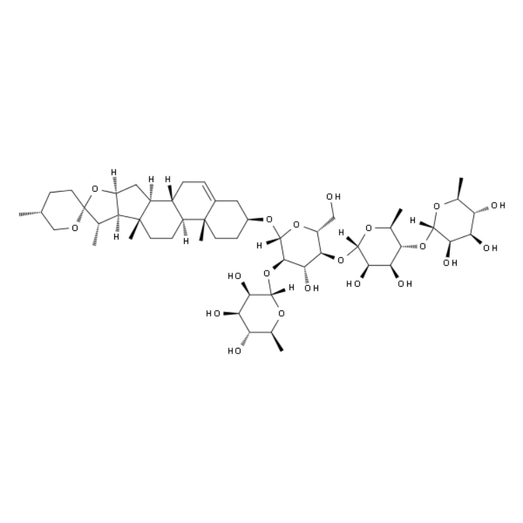
Polyphyllin II Provides a Model for Studying Cancer Cell Responses
MDA-MB-231 cells were used in the breast cancer studies with Polyphyllin II.
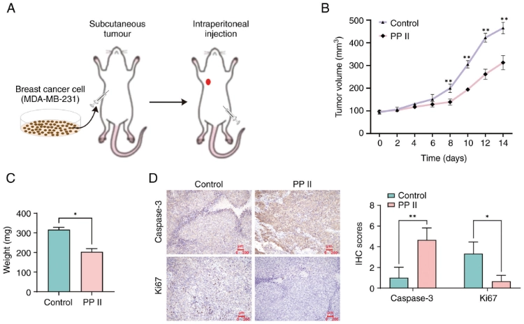
This is a good example of why one assay does not make a story.
A cell viability assay determines the number of viable cells after a treatment. It does not, however, explain why this number is decreased by the treatment.
Additional information on apoptotic cell populations can be obtained by flow cytometry measurements. Wound healing and invasion assays measure completely different characteristics of cancer cells, i.e. their motility and their change in invasive properties.
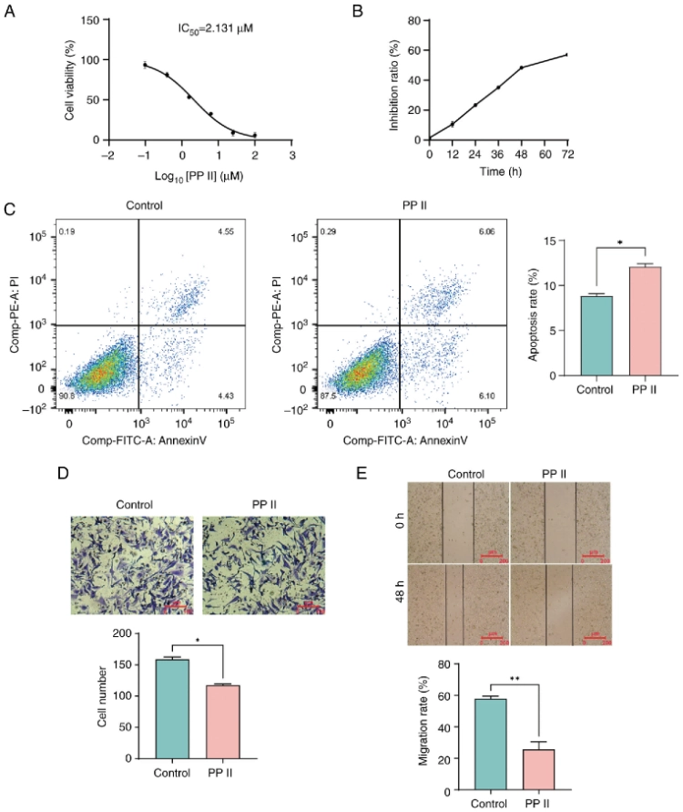
Instead of treating these results as interchangeable, it is more appropriate to read them together.
A low cell number could indicate slow growth or some other stress. However, when measuring apoptosis in several independent experiments, it is obvious what is going on.
Solarbio provides a wide range of small molecule compounds & related research products. Examples of compounds offered by Solarbio include Polyphyllin II for research use only.
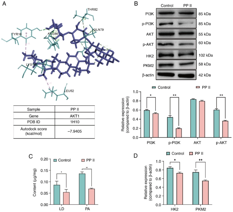
Why Do Apoptosis Experiments Need Multiple Detection Methods?
Apoptosis occurs over time. Early changes in signal transduction pathways, activation of caspases, changes in mitochondria and in the end the death of a cell do not occur simultaneously.
That is where a single assay can cause problems.
A viability assay measures the number of viable cells in a sample. However, it does not reveal the exact processes that occurred to the cells to reach this number.
An apoptosis experiment can include many different aspects depending on the question asked.
- Cell viability analysis
- Flow cytometry detection
- Caspase activity measurement
- Protein expression analysis
- Mitochondrial pathway evaluation
No need to add a plethora of methods to make an experiment look complete. You just need enough readouts to answer your question of interest and that is all.
How Can Researchers Improve Apoptosis Experiment Design?
Select Materials According to the Research Question
Start with the question, not the reagent catalog.
A cancer model, an immune study or a drug screening project all need apoptosis but differ in required setup.
A number of important matters should be established prior to the commencement of treatment:
- The cell model
- The compound being tested
- Treatment concentration and time
- The apoptosis markers to measure
- The final experimental purpose
The time of treatment is an easy parameter to overlook. A short treatment period may capture an early stage of a signaling pathway, whereas longer treatment periods may be required to observe obvious cell death.
Similarly, the optimal concentration must be found for a given assay. Concentrations that are too low might not give any signal, whereas too high concentrations might be so toxic that it masks the specific effect that one is trying to study.
More information about the research capabilities of Solarbio can be found in the Solarbio company profile.
Combine Cell Phenotype With Molecular Detection
The cells and the molecular data should correspond to each other.
So viability declines after treatment. What is causing this decline?
Apoptotic cell numbers can be determined by flow cytometry. Activation of the death cascade can be determined by measurement of caspases. Information on where a stress response is initiated can be obtained from mitochondrial or protein data.
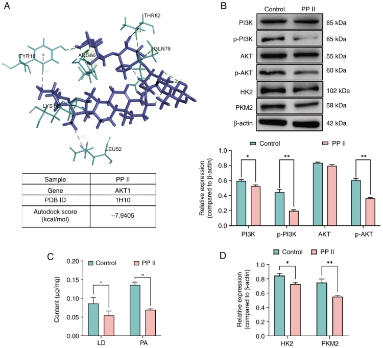
Basic consistency is important as well. This includes cell density, treatment time, concentration of test compounds, sampling time and others. The deviation in these parameters between different groups should not exceed the technical variation in order to avoid mixing up the biological variation with the technical variation.
Solarbio’s technical service resources are also available to help researchers select the right products as well as to plan experiments.
Conclusion
Apoptosis as a pathway is much more useful in practical lab work than just a means to count dead cells.
To study apoptosis, caspases, Bcl-2 family proteins, death receptors and mitochondria can serve as entry points. By using small molecule compounds to perturb one of these components, the subsequent effects can be analyzed.
When all parts of a research question, including the cell model, the treatment conditions and the detection methods, are well coordinated, the result is much easier to interpret. If the observed phenotype and the obtained molecular data fit well into the same story then also the underlying mechanism is much easier to support.
Solarbio offers a range of small molecule compounds & research reagents as well as related laboratory products for cell biology & molecular research studies. More information can be found on the Solarbio website, or users can contact Solarbio to discuss their experimental needs.
FAQ
Q1: What is the apoptosis pathway?
A1. Apoptosis is a controlled form of programmed cell death, which allows cells to die if they are damaged or are of no use anymore. This physiological process avoids inflammatory damage to surrounding tissues, and its dysregulation is closely related to tumour formation, tissue injury and degenerative diseases.
Q2: Which proteins are involved in apoptosis regulation?
A2: This includes proteins like the caspases, proteins of the Bcl-2 family, death receptors, Bax, Bak, cytochrome C and many more.
Q3: What is the difference between intrinsic and extrinsic apoptosis pathways?
A3: The extrinsic pathway starts with the death receptors on the surface of the cell, whereas the intrinsic pathway is initiated mainly by stress in the cell and is associated with the mitochondrial signaling pathway. The two pathways are not completely independent and have obvious signal crosstalk inside cells.
Q4: Why are small molecule compounds used in apoptosis studies?
A4: The researcher can select from a number of apoptosis‑related proteins for change, thereby facilitating the correlation of cellular responses with specific parts of the pathway. It should be cautioned that high compound concentrations may cause non‑specific toxicity, so proper concentration and treatment time need to be optimized in advance.
Q5: How is Polyphyllin II used in apoptosis research?
A5. Polyphyllin II is a natural steroidal saponin compound studied in cancer models. Using a combination of cell viability assays, flow cytometry, cell migration and invasion assays it is possible to investigate the effects of Polyphyllin II on cancer cells. Both in‑vitro cell experiments and in-vivo tumour-bearing mouse models are commonly applied to verify its pro-apoptotic anti-tumour effect.
 Consult
 Whatsapp
 Leave Message
 top