


Calcium Signaling Pathway
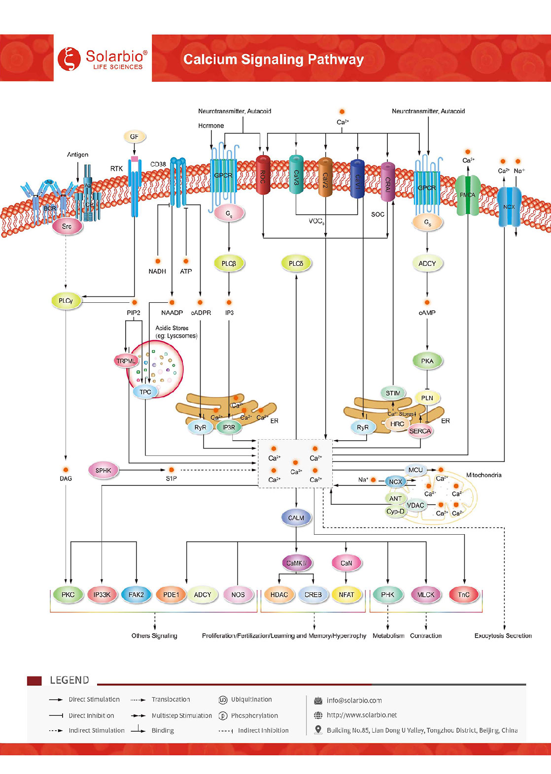
All physiological activities are regulated by Ca2+, and the basis of Ca2+ as a cellular messenger is the presence of a concentration gradient between cytosolic Ca2+ and intracellular stores or extracellular Ca2+. Ca2+ signaling is very flexible and at the same time tightly controlled. Ca2+ plays an important role in the regulation of cellular functions, such as nerve conduction, muscle contraction, morphological changes, nerve cell aging, secretion of gland cells and signal transmission of retinal cells. Many cellular functions depend on the extremely high difference between the intracellular and extracellular Ca2+ concentration. Once the difference between the intracellular and extracellular Ca2+ concentration is reduced, cellular functions will be damaged, and even cell death will be caused. Cytosolic Ca2+ comes from two sources: One is the extrinsic calcium entry pathway. Calcium channels are a class of transmembrane glycoproteins that form hydrophilic pores that approximate a funnel. They can be divided into three main categories: voltage-gated calcium channels (VOC), receptor-gated calcium channels (ROC), and store-controlled calcium channels (SOC). Among them, voltage-gated calcium channels catalyze rapid, highly selective Ca2+ influx into cells. The other major source is internal storage, which is mainly located in the endoplasmic reticulum/sarcoplasmic reticulum (ER/SR). There are two intracellular calcium release channels, IP3 and RYR receptor channels, which release calcium ions from ER/SR calcium stores. The main activator of these channels is Ca2+ itself, and this process of Ca2+ induced Ca2+ release is central to the Ca2+ signaling mechanism. Various second messengers or regulators also control Ca2+ release. IP3 regulates IP3R through the phospholipase C pathway. Cyclic ADP-ribose (cADPR) releases Ca2+ through RYRs. Nicotinic acid adenine dinucleotide phosphate (NAADP) stimulates Ca2+ release from acidic stores such as lysosomes and is a highly potent second messenger for calcium mobilization. Ca2+ released through the NAADP-sensitive mechanism may also feed back to RYR or IP3R. cADPR and NAADP are generated by CD38. The enzyme may be sensitive to cellular metabolism because ATP and NADH inhibit it. In addition, mitochondrial Ca2+ uptake is critical for cell survival and death. A calcium uniporter located in the inner mitochondrial membrane, responsible for mediating calcium uptake. This complex consists of two components, the selective calcium channel MCU and its regulator MICU1.
 Consult
 Whatsapp
 Leave Message
 top
top


