


Связанные новости


Cuproptosis Explained: A Practical Look at Copper-Driven Cell Death
Содержание
Cuproptosis has been talked about a lot since 2022. At first glance, it sounds like another cell death term that only matters to a small group of mechanism researchers. But once you look at how it works, it is easy to see why cancer biology, mitochondrial metabolism, inflammation research, and neurodegenerative disease studies all started paying attention to it.
The point is not simply that copper is toxic. Copper is a trace element, and cells do need it. The real problem starts when copper balance breaks. Too much free copper, especially Cu⁺, can push mitochondria into trouble. The cell then faces protein aggregation, metabolic stress, oxidative damage, and finally a special type of regulated cell death now known as Cuproptosis.
For researchers who work with cell models, this topic is not only about reading a pathway chart. It is also about compound selection, controls, purity, rescue experiments, and whether the same result can be repeated next week with another batch of reagent. That is where a supplier like Соларбио becomes relevant. Solarbio works in life science research tools, including small molecule compounds, biochemical reagents, ELISA kits, cell biology products, molecular biology reagents, antibodies, standards, and related products used in mechanism studies.
What Cuproptosis Means in Plain Lab Language
Cuproptosis is a copper-dependent regulated cell death pathway. It is different from apoptosis, ferroptosis, necroptosis, and classic oxidative cell damage. In Cuproptosis, the center of the story is the mitochondrion, especially the TCA cycle.
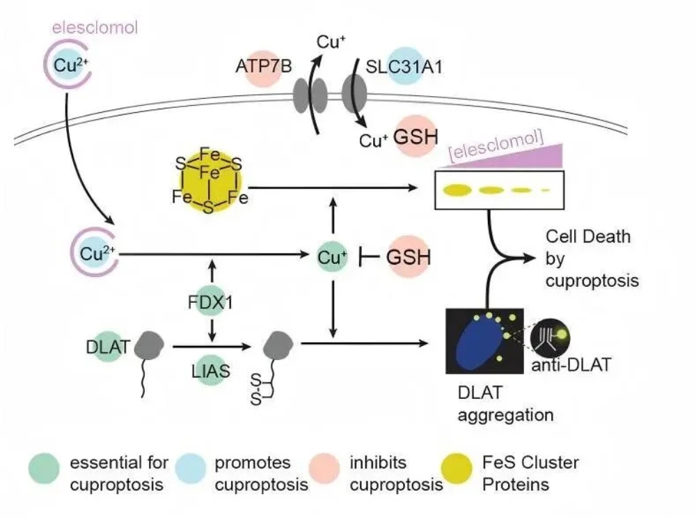
The key upstream factor is FDX1. FDX1 can reduce Cu²⁺ into Cu⁺. That sounds like a small chemical change, but in cells it matters a lot. Cu⁺ is more reactive and can make copper toxicity stronger. Once copper builds up, it binds to lipoylated proteins in the TCA cycle. DLAT and PDHA1 are two common examples mentioned in this field.
After that, the cell begins to lose control. These lipoylated proteins can form abnormal aggregates. As a secondary consequence, Fe-S proteins may also become destabilized. Mitochondrial function drops. Protein stress rises. This is not the usual caspase-driven apoptosis route. It is more like the cell’s energy engine becomes blocked by copper and protein stress at the same time.
This is why cuproptosis pathway research has become useful for people studying cancer metabolism, copper transport, mitochondrial injury, and drug resistance.
The Basic Mechanism Researchers Usually Watch
A Cuproptosis experiment often begins with copper homeostasis. Normal cells use transporters and binding proteins to keep copper under control. CTR1 helps bring copper into the cell. ATP7A/B helps remove copper. Metallothioneins can bind copper and lower free copper levels. When this system fails, copper does not stay quiet.
FDX1 then becomes very important. If FDX1 is active, Cu²⁺ can be reduced to Cu⁺. The toxic copper signal becomes stronger. The copper then binds lipoylated TCA cycle proteins, especially DLAT, and causes abnormal oligomerization. The combined effect—lipoylated protein aggregation together with secondary Fe-S protein instability—delivers a heavy hit on mitochondrial metabolism.
ROS can also increase during copper overload. That can damage DNA, membranes, and proteins. Copper has also been reported to interfere with UPS function in some contexts, which may exacerbate protein stress, but this is not a defining feature of cuproptosis. In actual experiments, these signals often appear together, so it is risky to say “this is Cuproptosis” based on only one marker.
A cleaner design usually checks several parts: copper treatment, copper ionophore induction, copper chelator rescue, FDX1 or DLAT change, mitochondrial morphology, and stress markers. This is also why stable life science research tools matter. If the reagent quality is not steady, the mechanism result becomes messy very quickly.

What Cells Look Like During Cuproptosis
Cuproptosis has some morphology that helps separate it from other cell death types.
The most obvious change is in mitochondria. Under transmission electron microscopy, researchers may see mitochondrial shrinkage, increased membrane density, and reduced or missing cristae. These signs fit the mechanism because Cuproptosis is closely tied to mitochondrial metabolism.
Other changes may also appear. Cells can shrink. The cell membrane may rupture. The endoplasmic reticulum can show damage. Chromatin may break, but classic apoptotic bodies are usually not the main feature. Caspase activation is not the central route either.
This does not mean morphology alone is enough. It is better to combine morphology with molecular data. For example, if copper treatment causes mitochondrial shrinkage, DLAT aggregation, FDX1-dependent sensitivity, and copper chelator rescue, the evidence becomes much stronger.
Key Genes and Pathways in Cuproptosis
Several genes are often used when researchers build a Cuproptosis-related panel.
FDX1 is usually placed at the center. LIAS and LIPT1 are tied to protein lipoylation. DLAT, DLD, PDHA1, and PDHB are connected with the TCA cycle. These genes support the copper-driven toxic process.
On the protective side, MTF1 helps regulate metallothionein expression and reduce free copper. GLS helps maintain metabolic balance.
Transporters are also worth checking. CTR1 brings copper in. ATP7A/B moves copper out. If CTR1 is high or ATP7A/B is low, cells may become more sensitive to copper accumulation. Nrf2 is another layer because it controls antioxidant response genes such as HO-1. When Nrf2 signaling is strong, cells may resist copper-related oxidative stress better.
In short, Cuproptosis is not one single switch. It is more like a copper-metabolism-mitochondria network. Changing one point may affect the whole result.
Why Cancer Researchers Care About Cuproptosis
Cancer cells often have unusual metal metabolism. Some tumor cells handle copper differently from normal cells. That gives researchers a possible opening. If a cancer cell is already sensitive to copper stress, pushing Cuproptosis may help weaken it.
AML is one disease area that gets attention. Some studies suggest that AML cells with lower heme biosynthesis enzyme expression, including HBE-related changes, may be more sensitive to copper accumulation. UM4118 has also been discussed in SF3B1-mutant and adverse-risk AML models.
In radioresistant tumors, MT1E and MT1X may bind copper and reduce copper toxicity. That can make tumor cells less sensitive to Cuproptosis. A copper ionophore may help reverse part of this resistance in some experimental designs.
Solid tumors are also being studied. Lung cancer, melanoma, and other models have all been connected with copper ionophore research. Still, the field should not be oversold. Copper can damage normal cells too. Dose, timing, cell type, transporter expression, and mitochondrial state all need to be checked carefully.
Cuproptosis in Inflammation, Brain Disease, and Copper Overload
Cuproptosis is not only a cancer topic.
In rheumatoid arthritis, serum copper can be higher, and genes such as LIAS and FDX1 may show abnormal expression. Copper-related synovial cell injury may add to inflammation and bone damage. In high glycolysis or hypoxic conditions, Cuproptosis may also be suppressed, which could allow immune cells to keep expanding.
In ulcerative colitis, copper accumulation in intestinal mucosa, increased DLAT oligomerization, and lower FDX1 expression have been discussed. If Cuproptosis damages the intestinal barrier, mucosal repair becomes harder.
Systemic lupus erythematosus has also been linked with LIAS-related Cuproptosis changes. The details still need more work, but the connection between copper balance and immune disorder is becoming harder to ignore.
Neurodegenerative disease is another area. In Alzheimer’s disease, copper can accumulate abnormally in the brain. That may speed up neuronal injury through mitochondrial dysfunction and protein stress. Parkinson’s disease may also involve copper-related neuronal damage.
Wilson disease is the classic example of copper overload. ATP7B mutation causes copper export failure. Copper then builds up in the liver, brain, and other organs. D-penicillamine and other copper chelation strategies can help reduce copper burden. This disease shows very clearly why copper control matters.
Small Molecule Compounds Used for Cuproptosis Work
In lab use, Cuproptosis-related compounds can be roughly divided into copper ionophores, copper chelators, and pathway regulators.
Copper ionophores help copper enter cells or move toward mitochondria. Disulfiram (ID2550) is known as an ALDH1 inhibitor. When it forms a complex with Cu²⁺, it can affect proteasome activity and has been used in cancer-related studies.
Zinc Pyrithione (IZ1140) is reported to have antifungal and antibacterial activity. It can also act as a copper ionophore, helping transport copper into cells. Because of that, it is often used as a tool compound in Cuproptosis research.
Elesclomol (IE0680) is a well-known copper ionophore. It can promote mitochondrial copper delivery and trigger oxidative stress. It is often used when researchers want to study copper-driven mitochondrial injury.
Cu(II)-Elesclomol (IYT2503) is the complex formed by Elesclomol and Cu²⁺. It can move copper into cancer cells, induce oxidative stress, promote lipoylated protein aggregation, and trigger Cuproptosis-related death. It can move copper into cancer cells, induce oxidative stress, promote lipoylated protein aggregation, and trigger Cuproptosis-related death. It has also been described as a weak DNA topoisomerase I inhibitor, so control design should be handled carefully.
UM4118 (IYT110497) is another tool compound for studying copper-dependent cell death. It can induce a mitochondria-based, non-classic cell death pattern and has shown selective toxicity in SF3B1-mutant and adverse-risk AML research, an effect that is copper-dependent.
Copper chelators work in the other direction. D-penicillamine (IP2220) can promote copper excretion. Emeramide(IE3810) is a thiol redox antioxidant and heavy metal chelator. TTM can bind copper strongly and is often discussed in Wilson disease and inflammation-related copper studies.
Practical Notes Before Running a Cuproptosis Experiment
A Cuproptosis experiment can go wrong for very simple reasons. The compound may not dissolve well. Copper concentration may be too high. Cells may already be stressed before treatment. A chelator rescue group may be missing. Or the researcher may only check ROS and then call it Cuproptosis, which is too thin.
A more practical setup should include untreated control, copper treatment, copper ionophore treatment, copper chelator rescue, mitochondrial observation, and at least one pathway-related marker such as FDX1 or DLAT. If possible, protein aggregation and Fe-S protein stability should also be checked.
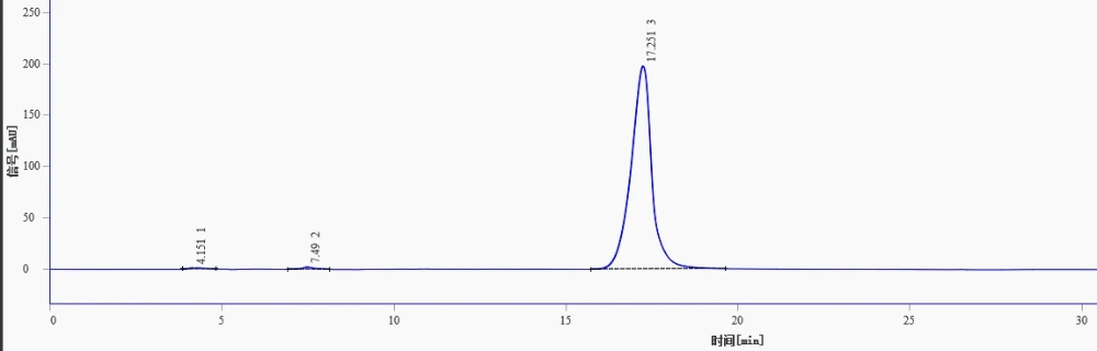
Compound purity also matters. The original product list includes Disulfiram, Zinc Pyrithione, Elesclomol, Cu(II)-Elesclomol, UM4118, D-penicillamine, and Emeramide, with related purity noted as ≥98%. For mechanism work, that kind of detail is not decoration. It affects whether the final data is believable.
For labs that are not sure which compound or assay fits their cell model, the technical service team can help with product selection and basic experiment matching.
Вывод
Cuproptosis gives researchers a useful way to study copper toxicity through mitochondria, lipoylated TCA cycle proteins, and secondary Fe-S protein instability, ROS stress, and protein aggregation. It has strong value in cancer metabolism, AML research, drug resistance, inflammatory disease, neurodegeneration, and copper overload disorders.
But this pathway is easy to oversimplify. Copper is not just poison. It is also a necessary metal element. Whether copper kills a cell depends on transporters, FDX1, mitochondrial state, protein lipoylation, antioxidant response, compound choice, and experimental dose.
For researchers planning Cuproptosis studies, Solarbio provides small molecule compounds, biochemical reagents, assay-related tools, and product support for early screening and follow-up validation. For product selection or project questions, researchers can связаться с Solarbio for more details.
Часто задаваемые вопросы
Q1: What is Cuproptosis?
A1: Cuproptosis is a copper-dependent regulated cell death pathway. It happens when copper disrupts mitochondrial metabolism, especially by binding lipoylated TCA cycle proteins and causing protein aggregation.
Q2: Is Cuproptosis the same as apoptosis?
A2: No. Apoptosis is usually caspase-related and may form apoptotic bodies. Cuproptosis is mainly linked to copper stress, mitochondrial damage, lipoylated protein aggregation, and secondary Fe-S protein instability.
Q3: Why is FDX1 important?
A3: FDX1 can reduce Cu²⁺ to Cu⁺. Cu⁺ is more toxic and can strengthen copper-induced cell death. FDX1 is also closely connected with the lipoylation-related part of the Cuproptosis mechanism.
Q4: What compounds can induce Cuproptosis?
A4: Common tool compounds include Disulfiram, Zinc Pyrithione, Elesclomol, Cu(II)-Elesclomol, and UM4118. They help increase copper delivery or copper-related mitochondrial stress.
Q5: What compounds can inhibit copper-related cell death?
A5: Copper chelators such as D-penicillamine, TTM, and Emeramide can reduce free copper or support copper clearance. They are often used in rescue experiments.
Q6: What genes are often checked in Cuproptosis research?
A6: Researchers often check FDX1, LIAS, LIPT1, DLAT, DLD, PDHA1, PDHB, MTF1, GLS, CTR1, ATP7A/B, Nrf2, and HO-1.
Q7: Can Cuproptosis be useful in cancer research?
A7: Yes. It is being studied in AML, lung cancer, melanoma, radioresistant tumors, and other cancer models. Some tumor cells may be more sensitive to copper stress because of abnormal copper metabolism.
Q8: Why does reagent quality matter in Cuproptosis experiments?
A8: Cuproptosis experiments are sensitive to compound purity, copper contamination, solvent choice, storage, and batch stability. Poor reagent quality can make the result look positive or negative for the wrong reason.


