



Noticias relacionadas
Double Immunodiffusion Test: Agar Gel Protocol, Results and Troubleshooting Guide
Tabla de Contenidos
The double immunodiffusion assay is a well-established technique in immunology laboratories. However,,obtaining crisp precipitin lines is often more challenging than anticipated. Even when antigens and antibodies are properly loaded onto an agarose gel plate, no precipitin reaction may be detected following incubation. Repeating the assay without troubleshooting critical experimental conditions only wastes valuable reagents.
The double immunodiffusion test, also known as the Ouchterlony assay, detects antigen–antibody interactions in agar gel. It requires relatively simple instrumentation and enables direct visualization of binding between soluble antigens and their cognate antibodies.
Laboratories working with immune serum, purified proteins, or antibody characterization may benefit from knowing the subtle details of this method to avoid excessive wasted time.
What Is the Double Immunodiffusion Test?
How Antigen-Antibody Precipitation Works
Double immunodiffusion relies on the migration of the soluble antigen and its cognate antibody within a gel matrix. The two components are loaded into discrete wells punched into an agarose plate.
Samples in solution diffuse into the gel. As the antigen and antibody migrate toward one another, the antigen encounters its cognate antibody, When the antigen and antibody concentrations are appropriately balanced,the two specifically recognize and associate to form immune complexes. As these complexes increase in size and lose insolubility, a distinct white precipitin line emerges.
This reaction typically occurs within the equivalence zone. At this optimal antigen‑to‑antibody ratio, large immune complexes assemble.
No labeling or fluorescence detection is required, as the precipitin-line can be directly visualized by the researcher. This technique is therefore especially suitable for basic immunology and preliminary antigen–antibody assays.
What Can Double Immunodiffusion Detect?
This assay enables identification of antigens and/or antibodies, as well as comparision of antibody activity via serial dilution of an antibody preparation.
It can also be applied to compare antigens derived from distinct samples. When multiple different antigen preparations are tested against the same antibody, the resulting precipitin lines reveal whether these antigens share recognizable antigenic determinants.
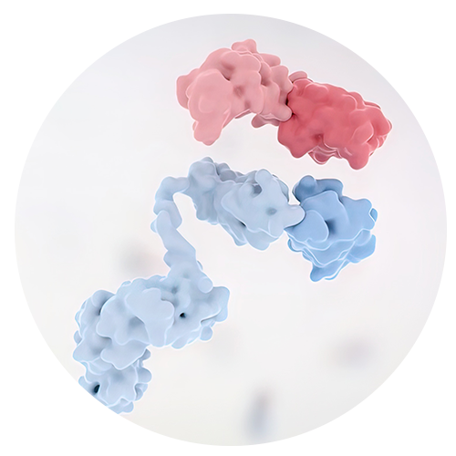
Antigen antibody binding is governed by antigen structure and the corresponding immune-mediated antibody recognition. In order to explore this relationship, one should start by familiarizing themselves with the biological molecule antibodies and how their structure determines immune recognition.
Double immunodiffusion serves primarily for qualitative and semi quantitative analysis; accordingly, it should not be confused with high sensitivity qualitative immunoassays.
Materials and Reagents for Double Immunodiffusion
Selecting Agarose and Buffer Solutions
Within the gel formulation., antigen and antibody samples are incorporated into a liquid medium. This medium typically consists of 1% agarose prepared in physiological saline or an appropriate buffer.
Agarose (Cat. No. A8201) is supplied by Solarbio for use in laboratory applications where a consistent gel matrix is required.
Make sure the agarose has completely dissolved before you pour it. Undissolved particles can interfere with the diffusion of the solutions and make faint precipitin lines hard to see.
Buffer selection is also critical. Physiological saline, phosphate buffer, and veronal buffer are compatible with suitable immunodiffusion systems. Normal saline (Cat. No. IN9004) can be used for gel preparation and sample dilutions.
Required supplies include Petri dishes, micropipettes, extra pipette tips, a gel punch, and a humidified incubation chamber.
Choosing Compatible Antigen and Antibody Samples
Specific binding must occur between the antigen and its cognate antibody. An unrelated antibody will not produce a precipitin line, even at high concentrations.
Bovine IgG dry powder (Cat. No. SP033) is used as an antigen in conjunction with Rabbit anti-bovine IgG (immune serum, Cat. No. SA233).
Human IgG dry powder (Cat. No. SP001) is an alternative antigen that can be paired with Goat anti-human IgG (immune serum, Cat. No. SA101).
Before starting, verify antibody specificity, sample concentrations, sample storage conditions, and whether the antigen in solution is still soluble.
How to Perform an Agar Gel Double Immunodiffusion Test
Preparing the Agarose Plate
Prepare a 1% agarose solution in physiological saline or another suitable buffer. Heat the mixture in a boiling water bath or microwave until the agarose dissolves completely.
Allow the solution to cool down to around 50–60°C and then pour it into a clean Petri dish. Aim for the gel thickness of 3–5 mm.
Do not disturb the plate until the agarose has solidified completely.
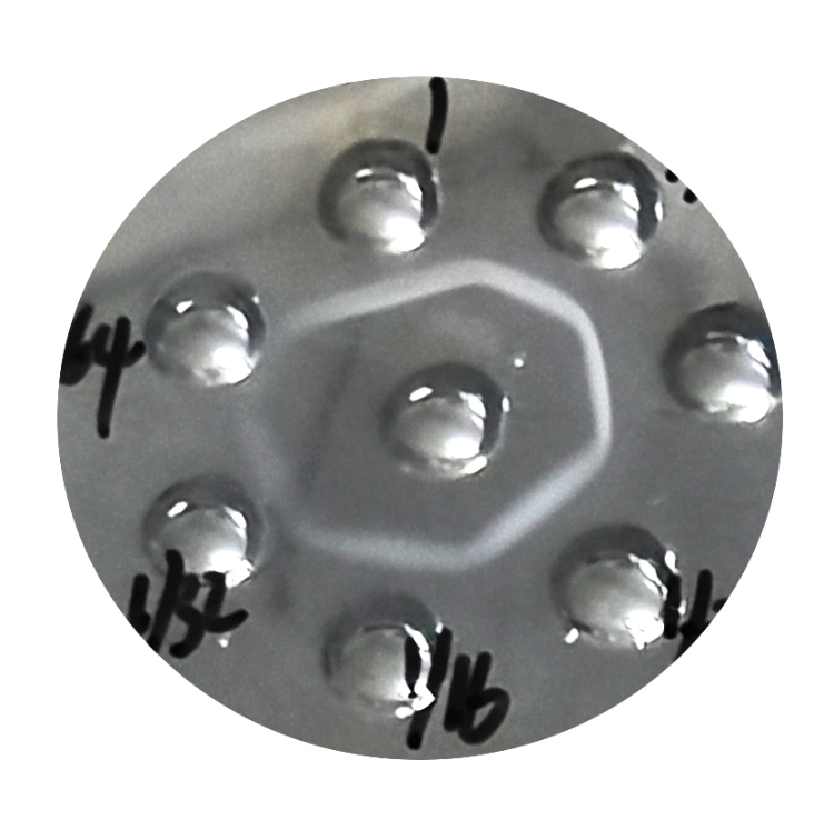
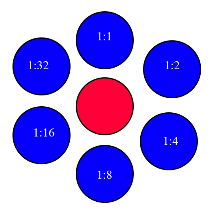
Cut wells using a gel punch. Most layouts have one central well surrounded by several outer wells to form a flower like pattern. The central well is approximately 3 mm in diameter, and outer wells are positioned 5–6 mm from the central well.
Carefully remove the agarose plugs from the wells. Irregular well edges could affect sample diffusion.
Verify that the bottom of each well is properly sealed to avoid sample leakage. The method of sealing will depend upon the material of the dish and direct flame should not be used to seal plastic dishes.
Loading Samples and Incubating the Plate
Before adding samples to the plate, mark the well positions on the underside of the plate.
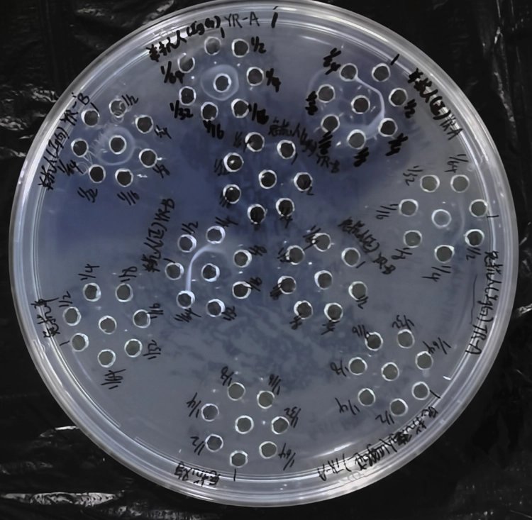
Load antigen and antibody solutions into the designated wells. Practically, a convenient experimental arrangement is to dispense one component at a fixed concentration into the central well, with serial dilutions of the second component loaded into the surrounding outer wells.
Use a micropipette to fill the gel wells. Take care not to spill the liquid on the gel surface.
A saline-only negative control is useful for unknown samples.
Cover the Petri dish and place it in the humidified incubation chamber. Incubate at about 37°C and inspect the plate periodically for 10-36 hours.
Examine the gel every 5–10 hours for the initial assay. Record the morphology and position of precipitin lines on the gel. Photographing the gel is strongly recommended.
The actual time for antigen-antibody interaction varies with multiple factors: antigen type, antibody concentration, diffusion properties, and gel matrix conditions.
Why Does Double Immunodiffusion Sometimes Fail?
No Visible Precipitin Line
The absence of a precipitin line does not rule out antigen–antibody reactivity.
When no precipitin line develops, one of the first parameters to check is whether the antigen or antibody concentration is too low to form detectable immune complexes.
Purified antibody solutions may start at a concentration of approximately 1 mg/ml or higher. However, the optimal concentration varies with the specific antibody and antigen being tested.
Excessively high antigen or antibody concentrations can also obscure the equivalence zone between wells. Rather than arbitrarily increasing concentration of both samples, prepare and test a dilution series.
It’s also important to check the sample identity. An antibody validated for Western blot or ELISA will not necessarily yield a visible precipitin line in agar gel.
Weak, Blurred, or Missing Lines After Incubation
A poorly prepared gel can make it difficult to visualize the reaction.
Uneven diffusion may occur if the agarose is not fully dissolved. Potential problems include damaged wells, sample leakage from wells, or uneven gel thickness.
Another parameter of importance is the humidity. When the gel dries locally, sample concentrations shift and the diffusion pattern can be affected. Keep the plate in a humidified chamber during the incubation.
A longer incubation time will not necessarily produce better visibility of precipitin lines. Extended incubation may also cause precipitin lines to disappear or turn diffuse.
If the initial run yields a distinct precipitin line at an earlier time point, use this time point for repeated assays.
How to Interpret Double Immunodiffusion Results
Understanding Precipitin Line Position
A precipitin line typically forms between the antigen and antibody wells, where their local concentrations reach the equivalence point.
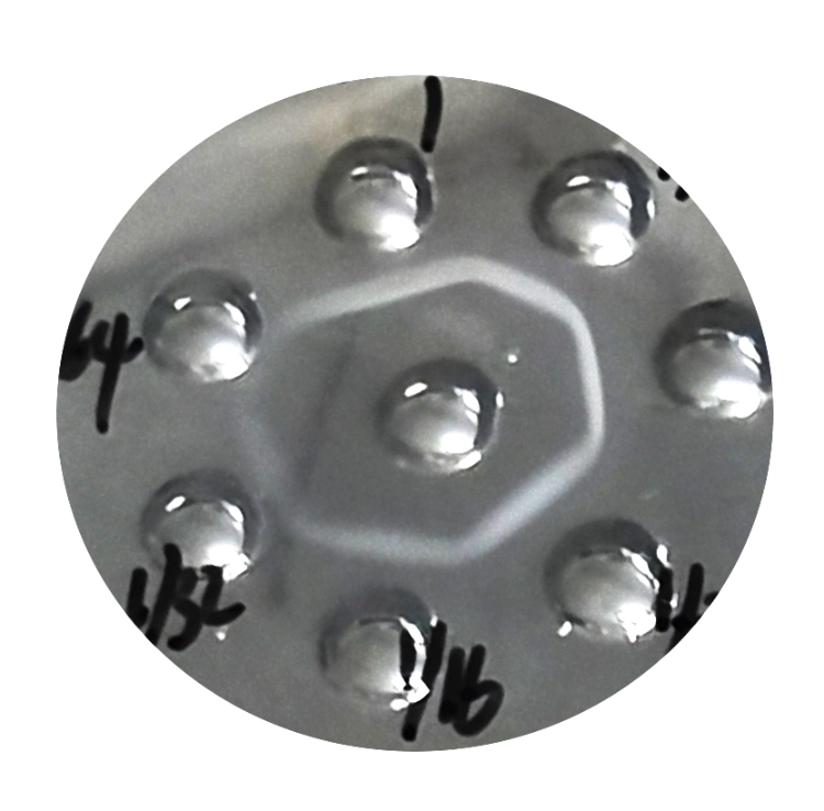
When the effective concentrations differ, the precipitin boundary will lie closer to the well holding the component at the lower concentration. Its position is also governed by the diffusion rates and other molecular characteristics of the reactants.
For comparison of multiple distinct antigens, the intersection pattern of precipitin lines provides particularly valuable information.
A fused continuous line indicates antigenic identity between the two samples. A spur signifies partial antigenic identity on the side where the spur forms. Crossing precipitin lines indicate non identical antigens.
These interpretive patterns are only reliable with appropriate reagents and well-defined reaction lines.
Double Immunodiffusion Compared With ELISA
Double immunodiffusion is used to detect precipitin lines or to study antigenic relationships. This technique is relatively low cost and requires no specialized detection equipment.
ELISA is more suitable for the determination of low concentrations of antigen and for obtaining quantitative results.
The guide published at Una guía para construir un sandwich ELISA serves as a useful reference for researchers choosing between Sandwich ELISA and alternative immunoassays, as it details the necessary reagents and key experimental stages for performing the assay.
The choice between experimental approaches should be driven by the research question, not by whether a method is newer or more established.
Conclusión
Classical double immunodiffusion is a technique for detecting antigen antibody complexes via precipitin formation and for comparing immune reactions. Most experimental failures stem from sample incompatibility, insufficient analyte concentrations, defective gels, poorly formed wells, or inappropriate diffusion conditions.
Starting with the suitable reagents and taking due care in sample handling is crucial to obtain reliable results. Solarbio offers a complete range of products including immune sera, IgG, agarose and more in the Solarbio Product Catalog.
For help selecting a compatible antigen-antibody pair or preparing materials for a new immunodiffusion experiment, contact Solarbio to discuss the required products and experimental conditions.
Preguntas frecuentes
Q1: How long does a double immunodiffusion test take?
A1: Incubation time for such an experiment is typically between 10 hours and 36 hours at about 37°C. This time depends on many factors, for example on the concentration of antigens/antibodies in the samples, on the composition of the gel and on the rate of diffusion of the samples through the gel. During the initial assay, check the gel at 5–10 hours intervals. Once you identify the time point at which precipitin lines appear most distinct, use that same endpoint for subsequent runs..
Q2: Why is there no precipitin line between the antigen and antibody wells?
A2. Possible reasons for no precipitin line: incompatibility antigen/antibody pairs, excessively low or excessively high sample concentrations, impaired diffusion, or damaged reagents. Run a known positive control first to validate the system. Prepare a dilution series for sample testing, rather than loading identical sample concentrations across multiple wells.
Q3: What agarose concentration is suitable for double immunodiffusion?
A3: 1% agarose gel is a good starting point. Make sure the agarose completely dissolves in the water. The final gel should be of even thickness, e.g. about 3-5 mm. The gel can differ between the various protein systems.
Q4: Can double immunodiffusion measure antibody titer?
A4. It is possible to establish a semi-quantitative estimate of antibody titer using serial dilution. The highest dilution at which a distinct precipitin line is visible under the selected assay conditions serves as the endpoint for this method. It does not have the same sensitivity and the same quantification range as ELISA.
Q5: How can I make faint precipitin lines easier to observe?
A5. Compatible antigen-antibody pairs should be used and their optimal concentrations determined. Sufficient moisture should be maintained in the gel and the wells should not be over-filled. The plate should be examined against a suitable contrast background and a photograph taken as the line develops. A dilution series is likely to be more effective than increasing the incubation time.
 Consultar
 Aplicación Whatsapp
 Dejar mensaje
 arriba