




Related news
Toll-like Receptors (TLRs): Immune Sentinels and Core Targets in Immune Regulation
Table of Contents
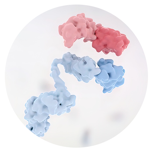
Toll-like receptors (TLRs) are widely investigated in inflammation and immunology research, especially in the fields of early infection, autoimmune diseases, tumor immunity and vaccine development.
When bacteria, viruses or damaged cells invade your body, the immune system needs to spot threats instantly. Serving as key pattern recognition receptors, TLRs act like immune sentinels—they help immune cells capture danger signals and trigger proper defense responses.
TLR signaling works in a sophisticated dual-regulation mode. Proper activation helps the body clear pathogens and build long-term immune memory. However, excessive activation will trigger persistent inflammation, tissue damage and immune disorders. As essential research tools for immune regulation, TLR-related experiments require well-designed models to avoid misleading results.
Solarbio offers a full portfolio of immunology research products, including small-molecule compounds, antibodies, ELISA kits, biochemical reagents and pathway-related products. High-quality reagents are the key to obtaining reliable data and minimizing background noise in all TLR studies.
What Are Toll-like Receptors?
TLRs Detect Danger Signals Early
TLRs are type I transmembrane proteins. They are highly expressed on innate immune cells such as macrophages and dendritic cells, as well as non-immune cells including epithelial cells and endothelial cells.
Their core function is pattern recognition. They can identify pathogen-associated molecular patterns (PAMPs) like bacterial lipopolysaccharide and viral double-stranded RNA. Besides, they also sense damage-associated molecular patterns (DAMPs) released by stressed or injured host cells.
Simply put, TLRs enable the body to quickly detect abnormal stimuli and launch targeted immune defenses.
Different TLRs Recognize Different Signals
Humans have 10 functional TLRs, which distribute in different subcellular locations: some localize on the cell surface, while others reside in intracellular endosomes.
Cell surface TLR1, TLR2, TLR4, TLR5 and TLR6 mainly recognize components from extracellular bacteria. Endosomal TLR3, TLR7, TLR8 and TLR9 specialize in detecting microbial nucleic acids, so they are widely studied in viral infection, antiviral immunity and nucleic acid-triggered inflammation.
When designing experiments, you need to clearly distinguish between bacterial challenge models and viral RNA stimulation models. Multiple factors including receptor location, ligand type, cell type and treatment duration should match your research goals. Common detection targets include activated TLR subtypes, downstream adaptor proteins and secreted cytokines.
How Do TLRs Regulate Immune Responses?
MyD88 and TRIF Are Two Main Routes
Once a TLR binds to its ligand, the intracellular TIR domain will kick off signal transduction with the help of adaptor proteins. Two classic pathways dominate this process: the myeloid differentiation primary response 88 (MyD88)-dependent pathway and the TIR-domain-containing adaptor-inducing interferon-β (TRIF)-dependent pathway.
These pathways further activate key transcription factors: nuclear factor-κB (NF-κB) and interferon regulatory factors (IRFs). Generally speaking, NF-κB drives the production of pro-inflammatory cytokines, while IRFs mainly mediate type I interferon responses. Still, the two pathways interact closely rather than working independently. For more credible results, we suggest testing multiple markers instead of a single indicator.
To explore interferon and cytokine functions, researchers often detect the expression of TNF-α, IL-6, IFN-α, IFN-β and their downstream molecules. Common experimental techniques include quantitative PCR, ELISA, Western blot, flow cytometry and tissue histological staining.
TLRs Start Innate Immunity
Innate immunity is the body’s rapid, non-specific defense line. Activated TLRs induce the release of pro-inflammatory cytokines, interferons, chemokines and antimicrobial peptides, recruiting neutrophils, macrophages and other immune cells to infection or injury sites.
Timely early immune responses effectively curb the spread of pathogens. But if inflammation lasts too long, the protective effect will reverse and cause tissue damage.
In TLR experiments, dose and treatment duration are two decisive factors. Even tiny changes in ligand concentration or treatment time will lead to totally different outcomes. You can select suitable reagents and assay kits from Solarbio’s product catalogue according to your research pathways, sample types and detection methods.
TLRs Link Innate and Adaptive Immunity
TLRs build a critical bridge between innate and adaptive immunity. When TLRs get activated, dendritic cells mature rapidly and enhance antigen presentation, which further activates T cells and B cells.
This is why TLR agonists are popular candidates for vaccine adjuvants. Their core value is to trigger powerful, long-lasting immune responses, instead of causing short-term excessive inflammation.
For vaccine-related TLR research, mainstream detection indicators include cytokine levels, antibody secretion, memory B cell formation, T cell activity and antigen presentation markers. A well-built experimental model is the foundation of trustworthy data.
Why Are TLRs Important Drug Discovery Targets?
Infection Research
In infectious disease research, TLR agonists are widely used to evaluate the body’s immune defense against various pathogens, especially viruses. TLR7/8 agonists show great potential in antiviral regulation, and are actively studied for treating chronic hepatitis B virus (HBV) infection to restore host antiviral ability.
Strong immune activation does not always mean good therapeutic effects. Some compounds cause severe non-specific inflammation and fail to act on specific signaling pathways. We recommend completing dose-response tests, setting sufficient controls and running repeated trials before carrying out large-scale formal experiments.
Autoimmune Disease Research
TLRs are closely linked to autoimmune disorders. Nucleic acid recognition by TLR7, TLR8 and TLR9 will drive aberrant immune activation. For this reason, TLR antagonists and pathway inhibitors have become promising therapeutic agents for systemic lupus erythematosus, rheumatoid arthritis and other related diseases.
A single cytokine test is far from enough to draw solid conclusions. Researchers also need to monitor immune cell counts, tissue inflammation, autoantibody levels and the activity of key signaling proteins. Autoimmune disease models are quite complex, so strict experimental controls are essential.
Cancer Immunology
TLRs play complicated roles in tumor immunity. Proper TLR agonists can activate antigen-presenting cells to trigger anti-tumor immunity and optimize the tumor microenvironment, making them hot research tools in cancer immunology.
Before using TLR ligands in tumor models, please clarify your research objectives first. Transient and moderate TLR activation helps fight against tumors, while long-term overactivation induces chronic inflammation and accelerates tumor growth. Currently, TLR ligands are widely applied to activate immune cells, inhibit tumor growth, induce cytokine secretion and boost the efficacy of combination therapy.
Product Examples Used in TLR Research
II0080 Imiquimod for TLR7-Related Models
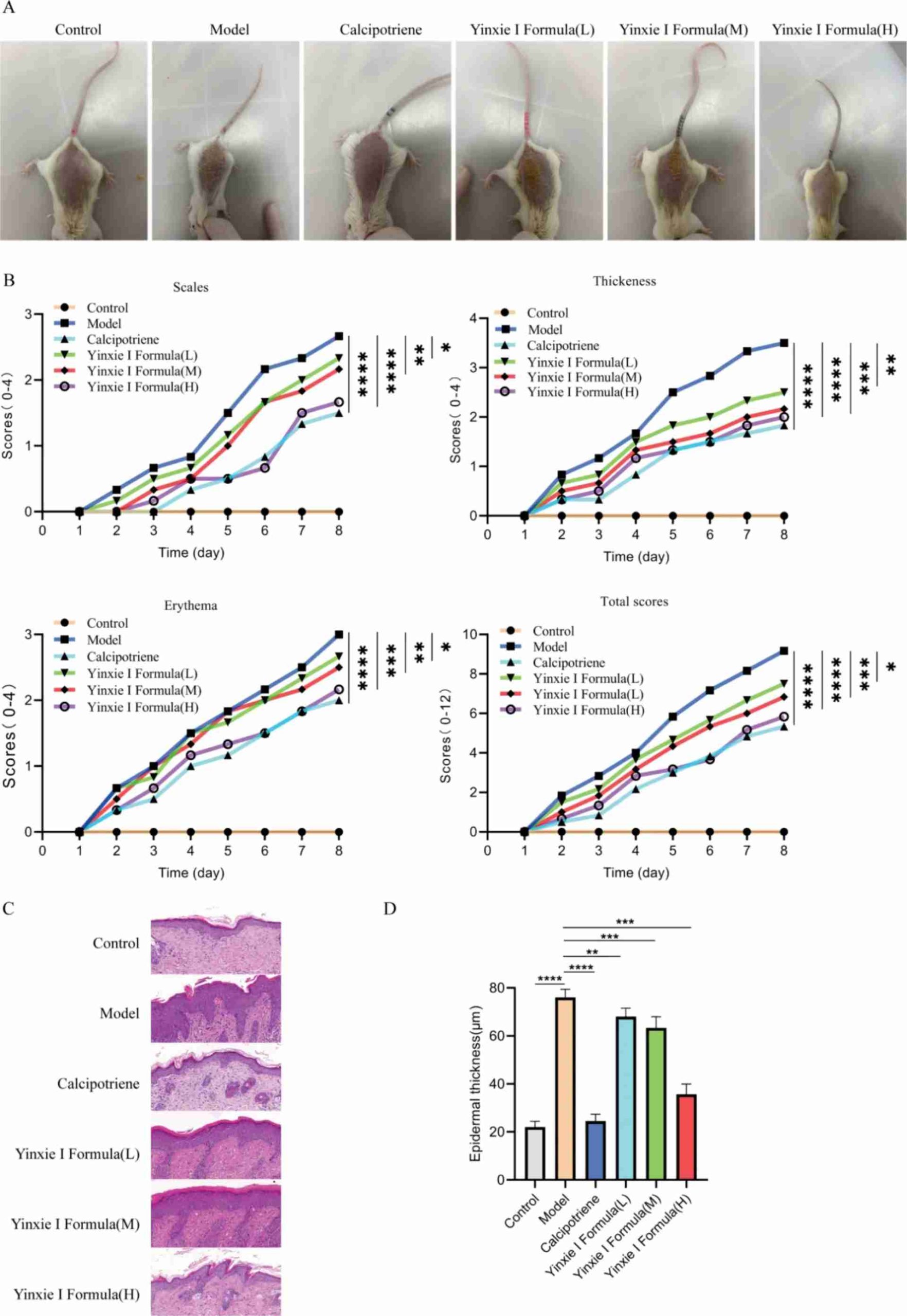
Small-molecule compounds like II0080 Imiquimod (IMQ) are classic workhorses for TLR research. As an immune response modifier and selective TLR7 agonist, imiquimod is widely used to activate immune responses and build classic inflammatory animal models, especially the IMQ-induced psoriasis-like skin inflammation model—a mainstream preclinical model for psoriasis and TLR7-mediated inflammatory mechanism research.
In published studies, 8-week-old male BALB/c mice (~25 g) received topical application of 62.5 mg 5% imiquimod cream daily for 7 consecutive days to build this stable disease model. After continuous IMQ stimulation, mice developed typical psoriasis symptoms including erythema, scales and skin thickening, which were confirmed by PASI scoring and HE tissue staining.
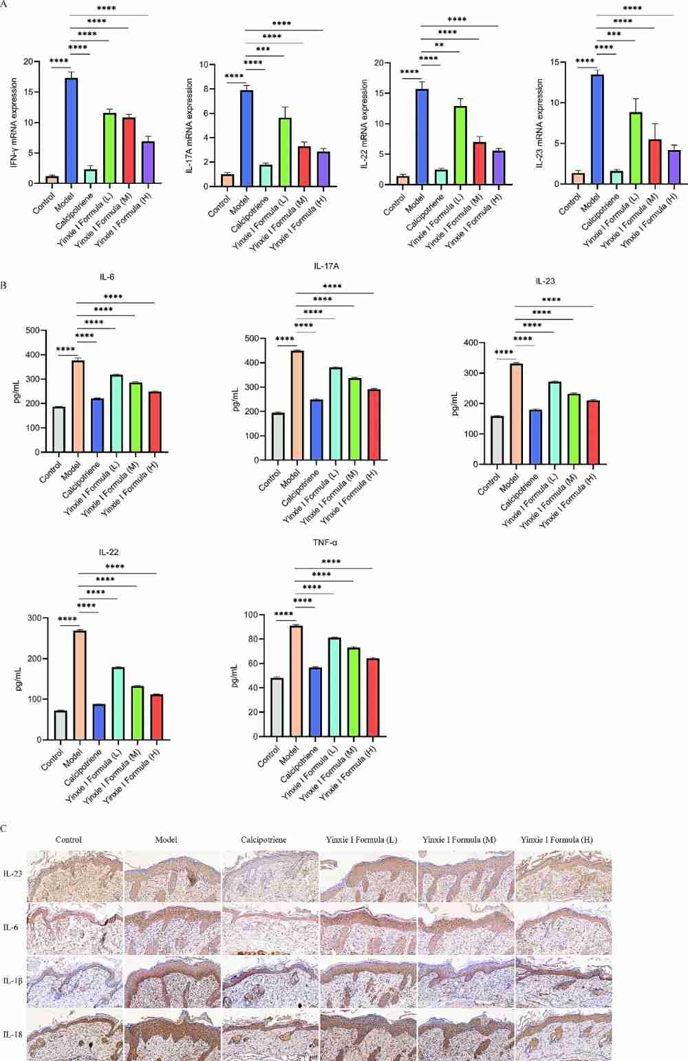
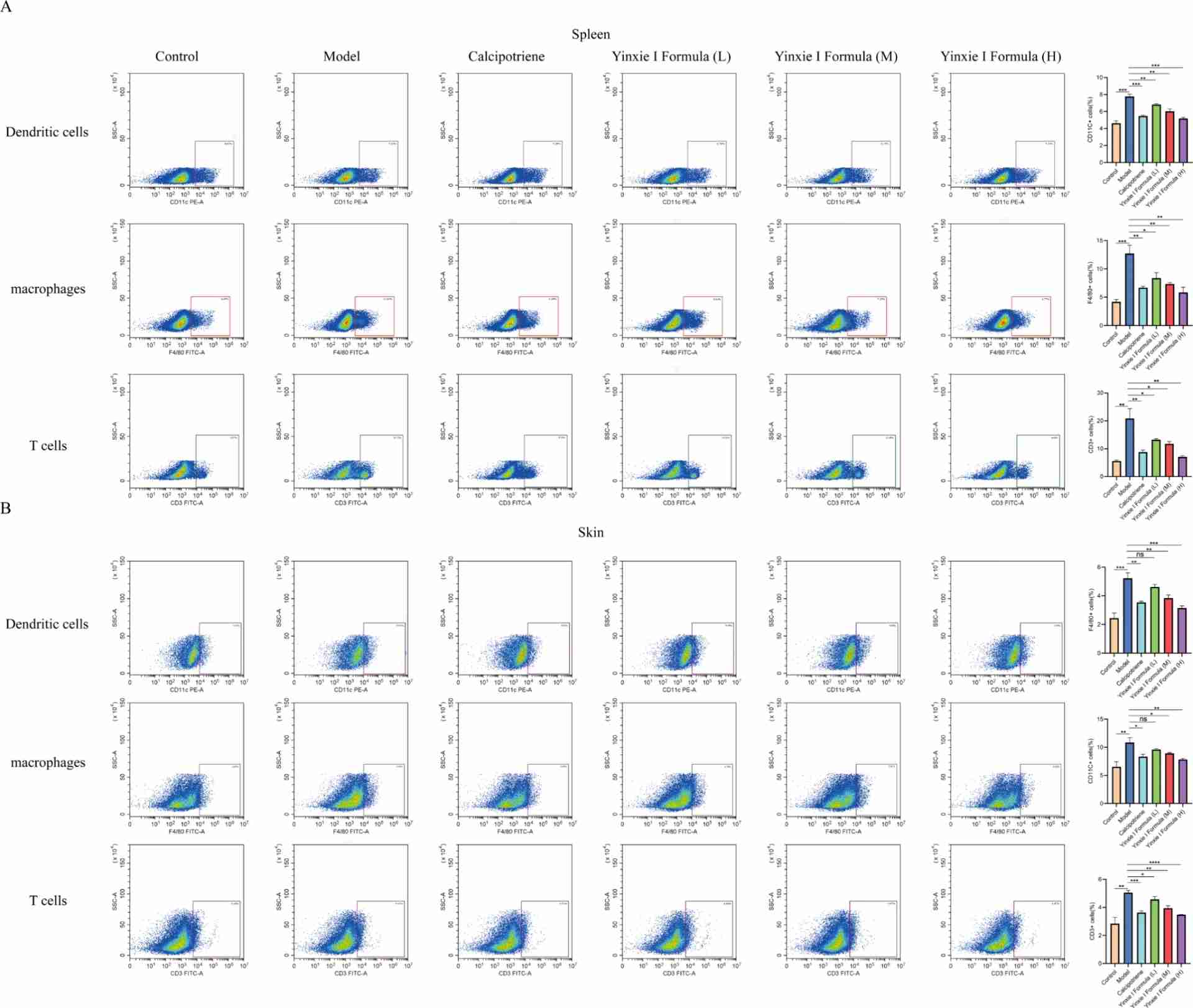
Activation of TLR7 by imiquimod significantly upregulates multiple pro-inflammatory factors such as IL-6, IL-17A, IL-22, IL-23 and TNF-α, with the IL-23/IL-17 axis acting as the key pathogenic pathway. Meanwhile, the quantities of dendritic cells, macrophages and T cells in skin and spleen also rise abnormally. Additionally, Western blot results verify that IMQ markedly increases the level of phosphorylated STAT3, revealing the crosstalk between TLR7 signaling and the STAT3 pathway.
Solarbio’s II0080 Imiquimod can effectively activate TLR7 and stably induce psoriasis-like inflammation. It is ideal for evaluating anti-inflammatory drugs and exploring TLR7-related inflammatory mechanisms. For reliable and repeatable data, please unify the administration dosage, application area, treatment duration and sample collection time during experiments.
IR0960 Resiquimod (R848) for TLR7/8 Stimulation
IR0960 Resiquimod (R848) is a potent dual TLR7/8 agonist, widely applied to activate immune cells and induce the secretion of cytokines including TNF-α, IL-6 and IFN-α. It is also a key stimulant for ELISpot assays targeting virus-specific memory B cells.
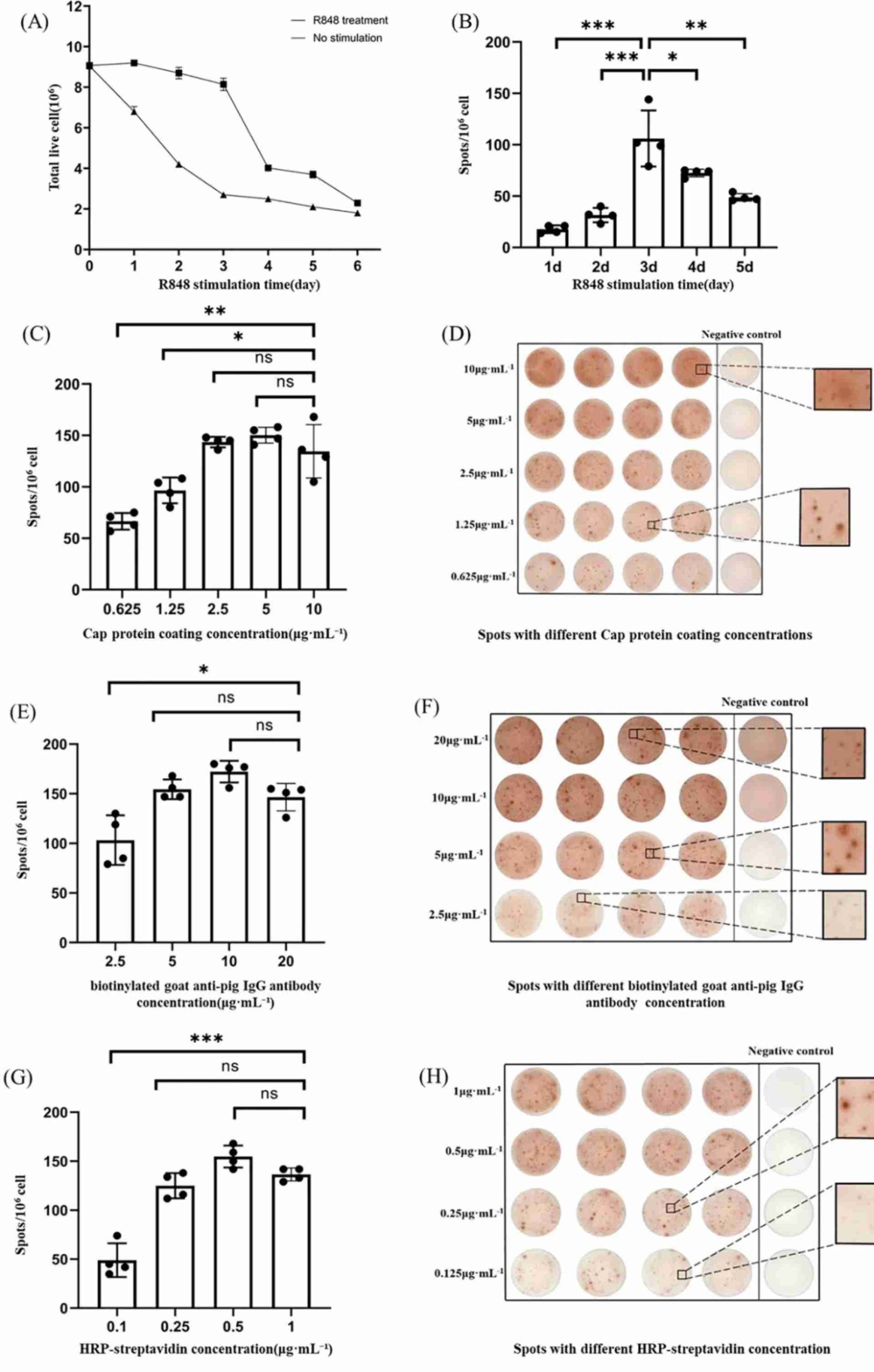
In typical PBMC-based experiments, the recommended working concentration of R848 is 1 µg/mL. Relevant methodological optimization verified that PBMCs stimulated with this concentration for 3 days achieved the highest activity of antigen-specific B cells while maintaining good cell viability.
For standard PCV2-specific memory B cell ELISpot detection, the matched optimal conditions are summarized as follows: PCV2 Cap protein coating at 1.25 µg/mL, biotinylated goat anti-pig IgG antibody at 5 µg/mL, and HRP-streptavidin at 0.25 µg/mL. These parameters deliver clear detection spots and low background signals, ensuring accurate and reliable assay results.
PBMC assays are prone to variability due to individual differences among donors. Key influencing factors include cell seeding density, culture duration and serum conditions. A complete experiment should set blank, solvent and stimulation control groups. If you are using R848 for the first time, we recommend conducting pilot tests in advance. Feel free to contact Solarbio’s technical team for professional support on experimental design and product selection.
How to Build a Better TLR Experiment
Match the Ligand, Cell Type, and Readout
Your experimental model must fit your research goals perfectly. For TLR7/8 studies focusing on cytokine production, PBMCs are a better choice than unrelated cell lines. When exploring skin inflammation, combining tissue scoring, cytokine detection and pathological analysis will bring you more comprehensive data.
A common mistake in experiment design is picking ligands first and then adapting the model reluctantly. The smarter way is to define your research hypothesis first, then select matching receptors, ligands, samples, treatment time and detection indicators.
Control Dose, Time, and Reagent Consistency
TLR activation triggers transient biological changes. Typically, the upregulation of target gene mRNA appears earlier than protein secretion. Cytokine protein detection requires longer cell culture, and obvious tissue changes usually take several days to develop.
Choosing the right compound dose is critical. High doses produce clear signals but may cause cellular stress and toxic interference; low doses better simulate natural immune responses yet often deliver weak signals. It is necessary to confirm the effective dose range beforehand, especially when using compounds on new cell lines or animal models.
Apart from experiment design, reagent quality directly impacts data stability. Batch differences may lead to fluctuating cytokine levels and high background staining. Founded in 2004, Solarbio is a professional supplier of reagents for immunology, cell biology, molecular biology and biochemistry. We have obtained ISO 9001, ISO 13485, ISO 14001 and ISO 45001 certifications. Learn more about us on our official “About Solarbio” page.
Conclusion
TLRs are indispensable regulators of immune activities. They recognize pathogen and danger signals, activate innate immunity and connect adaptive immunity. Therefore, they have become core research targets for infectious diseases, autoimmune disorders, tumor immunology and vaccine adjuvant development.
TLR signaling is never a simple on-off switch. Reliable experimental results rely on many factors: TLR subtypes, ligand features, compound concentration, cell/animal models, treatment duration and detection methods. Successful TLR experiments require careful planning, stable reagents and standard controls.
II0080 Imiquimod and IR0960 Resiquimod (R848) are two widely recognized small-molecule tools for TLR research. To get ideal data, please match them with suitable experimental models and detection systems. If you have demands for product selection or experiment protocols, welcome to contact us with your target receptor, sample type and detection requirements.
FAQ
Q1: What are Toll-like receptors in simple terms?
A1: Toll-like receptors are immune receptors that spot pathogen and danger signals, and trigger immune responses to defend against bacteria, viruses and damaged cells.
Q2: Why are TLRs called immune sentinels?
A2: As the frontline pattern recognition receptors in the body, TLRs quickly detect PAMPs from pathogens and DAMPs from damaged cells, and start downstream immune signaling. That’s why we call them immune sentinels.
Q3: Which TLRs are there that are linked to viral nucleic acid sensing?
A3: TLR3, TLR7, TLR8 and TLR9 are located inside cells and mainly responsible for sensing viral nucleic acids. They are core research objects in antiviral immunity.
Q4: What is II0080 Imiquimod used for in TLR studies?
A4: II0080 Imiquimod is a classic TLR7 agonist. It is widely used to build immune activation models, as well as psoriasis-like skin inflammation and other inflammatory models.
Q5: What is IR0960 Resiquimod (R848) used for?
A5: IR0960 Resiquimod (R848) is a dual TLR7/8 agonist, which can induce the secretion of TNF-α, IL-6, IFN-α and other cytokines in immune cells.
Q6: What do you check before starting a TLR experiment?
A6. You need to confirm ligand purity, storage conditions, solvent formula, dose range, treatment duration, experimental controls, sample type and detection methods in advance.
 Consult
 Whatsapp
 Leave Message
 top
top