




ข่าวที่เกี่ยวข้อง
วิธีการตรวจจับอะพอพโทซิส: การเลือกวิธีการทดสอบ เคล็ดลับเชิงปฏิบัติ และชุดอุปกรณ์ที่แนะนำ
ตารางเนื้อหา
อะพอพโทซิสเป็นกระบวนการตายของเซลล์แบบมีโปรแกรมควบคุมขั้นพื้นฐาน มีความสำคัญอย่างยิ่งต่อการเจริญเติบโต ความสมดุลของเนื้อเยื่อ การพัฒนาของโรค และผลของยา กระบวนการนี้มีความเกี่ยวข้องอย่างมากกับการศึกษาโรคมะเร็ง ปัญหาของระบบภูมิคุ้มกัน ชีววิทยาของระบบประสาท และการพัฒนายา
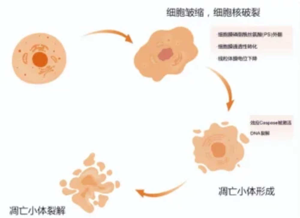
อย่างไรก็ตาม ในการทดลองจริง ความยากลำบากมักไม่ได้อยู่ที่การวัดอะพอพโทซิสโดยตรง ปัญหาที่ยากกว่าคือการเลือกเทคนิคที่เหมาะสมกับวัสดุ เวลา และเป้าหมายการศึกษา คุณอาจต้องการแยกแยะอะพอพโทซิสในระยะเริ่มต้นออกจากระยะท้าย หรือคุณอาจต้องการแยกแยะอะพอพโทซิสออกจากเนื้อตาย นอกจากนี้ คุณอาจต้องการตรวจสอบว่าความเสียหายของไมโทคอนเดรีย การกระตุ้นของเอนไซม์แคสเปส หรือการแตกหักของดีเอ็นเอเป็นสาเหตุของผลการวิจัยหรือไม่
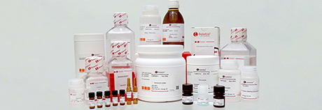
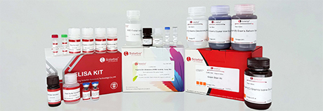
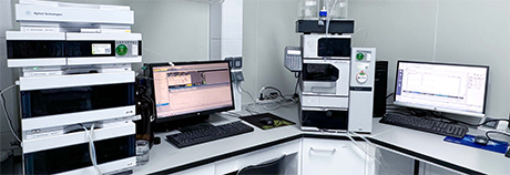
สำหรับห้องปฏิบัติการที่ต้องการขั้นตอนการทดสอบที่เชื่อถือได้ แทนการซื้อแบบครั้งเดียวจบ ปักกิ่ง Solarbio Science & บริษัท เทคโนโลยี จำกัด นำเสนอทางเลือกที่เหมาะสม บริษัทนี้ก่อตั้งขึ้นในปี 2547 และผลิตและส่งมอบผลิตภัณฑ์และบริการต่างๆ เครื่องมือวิทยาศาสตร์ชีวภาพ เช่น ชุดทดสอบเซลล์ ชุดทดสอบสารเคมี สารละลายสี แอนติบอดี สารอ้างอิง และสารโมเลกุลขนาดเล็ก มาตรฐานของบริษัท ได้แก่ ISO 9001, ISO 13485, ISO 14001 และ ISO 45001 นอกจากนี้ยังมีพื้นที่จัดเก็บข้อมูลที่ครอบคลุมและ ระบบช่วยเหลือ. ในการศึกษาเกี่ยวกับการตายของเซลล์ ความน่าเชื่อถือเช่นนี้มีความสำคัญอย่างยิ่ง สัญญาณเรืองแสงที่คงที่ ผลลัพธ์สีที่คงที่ และการตั้งค่าการทดสอบที่สม่ำเสมอ ล้วนส่งผลต่อคุณภาพของผลลัพธ์เป็นอย่างมาก
เทคนิคการตรวจจับอะพอพโทซิสโดยทั่วไปแบ่งออกได้เป็น 3 กลุ่ม:
การทดสอบทางชีววิทยาเชิงฟังก์ชัน
การตรวจวิเคราะห์ตัวบ่งชี้ทางชีวเคมี
วิธีการสังเกตทางสัณฐานวิทยา
แต่ละกลุ่มจะแก้ปัญหาเฉพาะด้านในห้องปฏิบัติการ ดังนั้น ผลลัพธ์ที่ดีที่สุดจึงมักเกิดขึ้นจากการใช้วิธีการหลายวิธีร่วมกัน
การทดสอบการทำงานเพื่อศึกษาภาวะอะพอพโทซิสระยะเริ่มต้นและภาวะเครียดของไมโทคอนเดรีย
การทดสอบการทำงานมีประโยชน์อย่างยิ่งเมื่อต้องการตรวจจับภาวะอะพอพโทซิสก่อนที่จะเกิดความเสียหายทางโครงสร้างอย่างร้ายแรง เทคนิคเหล่านี้เหมาะสำหรับการตรวจสอบเบื้องต้น การทดสอบยา และการวิจัยเกี่ยวกับความเสียหายของไมโทคอนเดรีย
การทดสอบศักย์เยื่อหุ้มไมโตคอนเดรีย
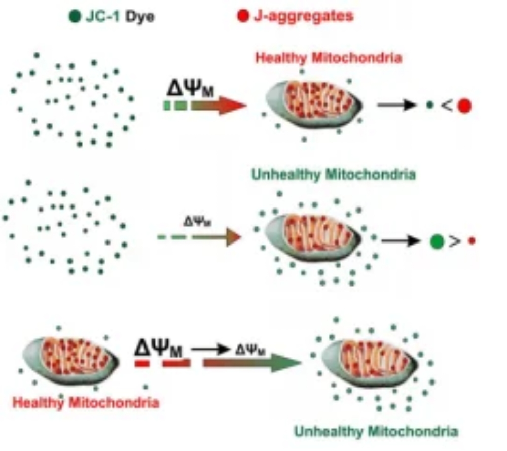
การลดลงของศักยภาพเยื่อหุ้มไมโทคอนเดรียเป็นหนึ่งในเหตุการณ์เริ่มต้นที่เกี่ยวข้องกับกระบวนการอะพอพโทซิส เมื่อความเสถียรของไมโทคอนเดรียเปลี่ยนแปลงไป ศักยภาพของเยื่อหุ้ม หรือที่เรียกว่า ΔΨm ก็จะลดลง
JC-1 และ JC-10 ทำหน้าที่เป็นตัวบ่งชี้เรืองแสงทั่วไปสำหรับงานนี้ ในไมโทคอนเดรียที่แข็งแรง JC-1 จะสร้างกลุ่มเรืองแสงสีแดง เมื่อศักยภาพลดลง มันจะคงอยู่เป็นหน่วยเดี่ยวเรืองแสงสีเขียว การเปลี่ยนแปลงจากสีแดงเป็นสีเขียวนี้เป็นสัญญาณที่ชัดเจนของการเสื่อมสภาพของไมโทคอนเดรีย
| หมายเลขแคตตาล็อก | ชื่อสินค้า | ความแม่นยำ | ความไว | ความเป็นพิษต่อเซลล์ | ใช้งานง่าย |
|---|---|---|---|---|---|
| ซีเอ1310 | ชุดตรวจวัดศักย์เยื่อหุ้มไมโตคอนเดรีย JC-10 สีเขียว/แดง | ***** | ***** | ต่ำมาก | ***** |
| M8650 | ชุดตรวจวัดศักย์เยื่อหุ้มไมโตคอนเดรีย JC-1 สีเขียว/แดง | **** | **** | ต่ำมาก | ***** |
| ซีเอ3660 | ชุดตรวจวัดศักย์เยื่อหุ้มไมโตคอนเดรีย (TMRE) สีส้มแดง | *** | *** | ต่ำมาก | ***** |
| ซีเอ3670 | ชุดตรวจวัดศักย์เยื่อหุ้มไมโตคอนเดรีย โรดามีน 123 สีเหลืองเขียว | ** | ** | ต่ำ | **** |
| เจ8030 | JC-1 โพรบวัดศักย์เยื่อหุ้มไมโทคอนเดรีย สีเขียว/แดง | **** | **** | ต่ำมาก | ***** |
| เจ8050 | JC-10 โพรบวัดศักย์เยื่อหุ้มไมโทคอนเดรีย สีเขียว/แดง | ***** | ***** | ต่ำมาก | ***** |
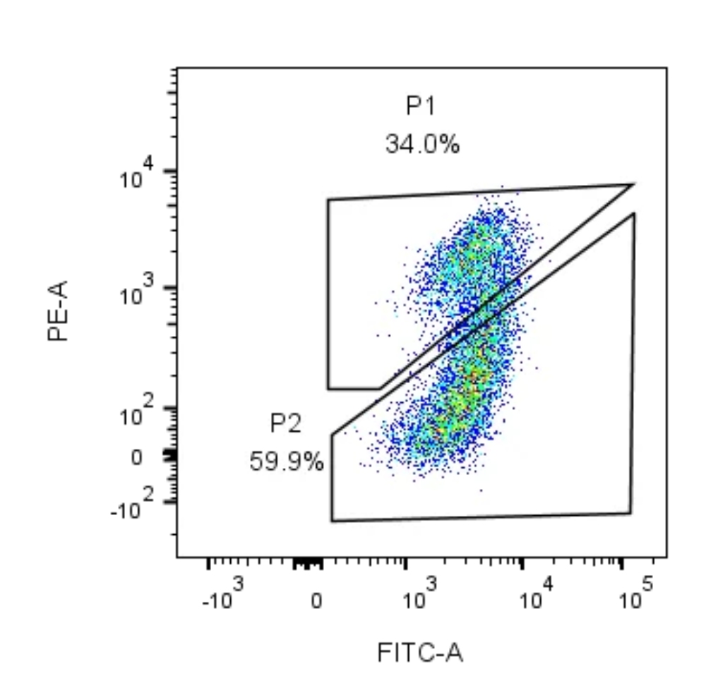
เทคนิคนี้ถือเป็นตัวเลือกที่เชื่อถือได้เมื่อประเมินภาวะอะพอพโทซิสของไมโทคอนเดรีย ความเสียหายจากอนุมูลอิสระ หรือความเครียดเบื้องต้นที่เกิดจากยา
การตรวจจับภาวะอะพอพโทซิสระยะเริ่มต้นและระยะท้ายโดยใช้แอนเน็กซินวี
หากคุณต้องการแยกเซลล์ที่มีชีวิต เซลล์ที่กำลังจะตาย และเซลล์ที่กำลังจะตาย การย้อมสีด้วยแอนเน็กซิน V มักเป็นตัวเลือกที่ดีที่สุด
ในเซลล์ที่แข็งแรง ฟอสฟาติดิลเซอรีนจะอยู่ด้านในของเยื่อหุ้มเซลล์ ในระยะเริ่มต้นของการเกิดอะพอพโทซิส ฟอสฟาติดิลเซอรีนจะเคลื่อนตัวไปยังพื้นผิวด้านนอก แอนเน็กซิน V จะเกาะติดกับฟอสฟาติดิลเซอรีนที่ปรากฏออกมานี้ เมื่อใช้ร่วมกับ PI หรือ 7-AAD คุณสามารถตรวจสอบความแข็งแรงของเยื่อหุ้มเซลล์ได้เช่นกัน
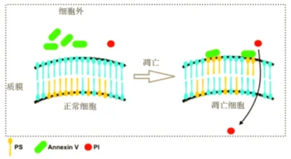
การตีความโดยทั่วไป:
แอนเน็กซิน วี-/พีไอ-: เซลล์ที่มีชีวิต
แอนเน็กซิน V+/PI-: เซลล์ที่อยู่ในระยะเริ่มตายแบบอะพอพโทซิส
แอนเน็กซิน V+/PI+: เซลล์ที่อยู่ในระยะอะพอพโทซิสตอนปลายหรือเซลล์ที่ตายแล้ว
แอนเน็กซิน V-/PI+: เซลล์ที่มีเยื่อหุ้มเซลล์เสียหาย
| หมายเลขแคตตาล็อก | ชื่อสินค้า |
|---|---|
| CA1020 | ชุดตรวจจับอะพอพโทซิส Annexin V-FITC/PI |
| CA1040 | ชุดตรวจจับอะพอพโทซิส Annexin V Alexa Fluor 488/PI |
| CA1050 | ชุดตรวจจับอะพอพโทซิส Annexin V Alexa Fluor 647/PI |
| ซีเอ3580 | ชุดตรวจจับอะพอพโทซิส Annexin V-APC/PI |
| CA1030 | ชุดตรวจจับอะพอพโทซิส Annexin V PE/7-AAD |
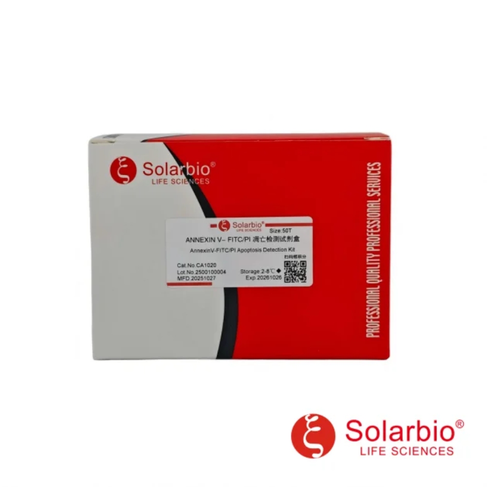
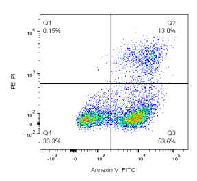
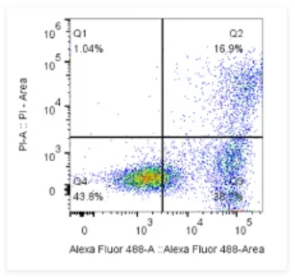
การทดสอบนี้เหมาะสมอย่างยิ่งกับระบบโฟลว์ไซโตเมทรี นักวิจัยมักเลือกใช้การทดสอบนี้เพื่อตรวจสอบจำนวนอะพอพโทซิส
รูพรุนการเปลี่ยนแปลงสภาพการซึมผ่านของไมโตคอนเดรียและการสะสมแคลเซียมมากเกินไป
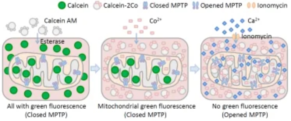
อะพอพโทซิสบางประเภทมีปัญหาเกี่ยวกับไมโทคอนเดรียอย่างรุนแรง โดยการเปิดของรูพรุนการเปลี่ยนแปลงสภาพความซึมผ่านของไมโทคอนเดรียและปริมาณแคลเซียมที่มากเกินไปภายในเซลล์เป็นข้อมูลสำคัญที่อธิบายกลไกต่างๆ
การเปิดของ MPTP อาจเกิดขึ้นได้จากภาวะแคลเซียมเกิน ภาวะเครียดออกซิเดชัน การเปลี่ยนแปลงของกลูตาไธโอน การหลุดออกของไซโตโครมซี และการสูญเสียศักยภาพของเยื่อหุ้มไมโทคอนเดรีย
| หมายเลขแคตตาล็อก | ชื่อสินค้า |
|---|---|
| ซีเอ3650 | ชุดตรวจจับรูพรุนการเปลี่ยนแปลงสภาพการซึมผ่านของไมโทคอนเดรีย สีเขียว |
แคลเซียมส่วนเกินยังสามารถเร่งกระบวนการอะพอพโทซิส (การตายของเซลล์) ได้อีกด้วย เพื่อตรวจสอบเรื่องนี้ คุณอาจใช้สารเรืองแสงที่บ่งชี้แคลเซียม เช่น Fluo-3, Fluo-4 และ Fluo-8
| หมายเลขแคตตาล็อก | ชื่อสินค้า |
|---|---|
| ซีเอ1180 | ชุดตรวจจับไอออนแคลเซียม Fluo-3 AM |
| ซีเอ1190 | ชุดตรวจจับไอออนแคลเซียม Fluo-4 AM |
| เอฟ8841 | Fluo-3 AM Calcium Probe ความเข้มข้น 5 mM ใน DMSO ปราศจากน้ำ |
| เอฟ8840 | โพรบวัดแคลเซียม Fluo-3 AM |
| เอฟ8501 | Fluo-4 AM Calcium Probe ความเข้มข้น 5 mM ใน DMSO ปราศจากน้ำ |
| เอฟ8500 | โพรบวัดแคลเซียม Fluo-4 AM |
| เอฟ8960 | โพรบไอออนแคลเซียม Fluo-8 AM |
เครื่องมือเหล่านี้มีประโยชน์ในการเชื่อมโยงกระบวนการอะพอพโทซิสกับความเสียหายของไมโทคอนเดรีย เส้นทางความเครียด หรือการเปลี่ยนแปลงของไอออน
ตัวบ่งชี้ทางชีวเคมีที่ช่วยเสริมความแข็งแกร่งให้กับข้อสรุปของคุณ
การทดสอบการทำงานแสดงให้เห็นถึงความเสียหายเบื้องต้น แต่ก็ไม่ได้ยืนยันลำดับการเกิดอะพอพโทซิสอย่างสมบูรณ์เสมอไป ตัวบ่งชี้ทางชีวเคมีจึงเข้ามาเติมเต็มช่องว่างนี้
การตรวจจับกิจกรรมของเอนไซม์ Caspase-3
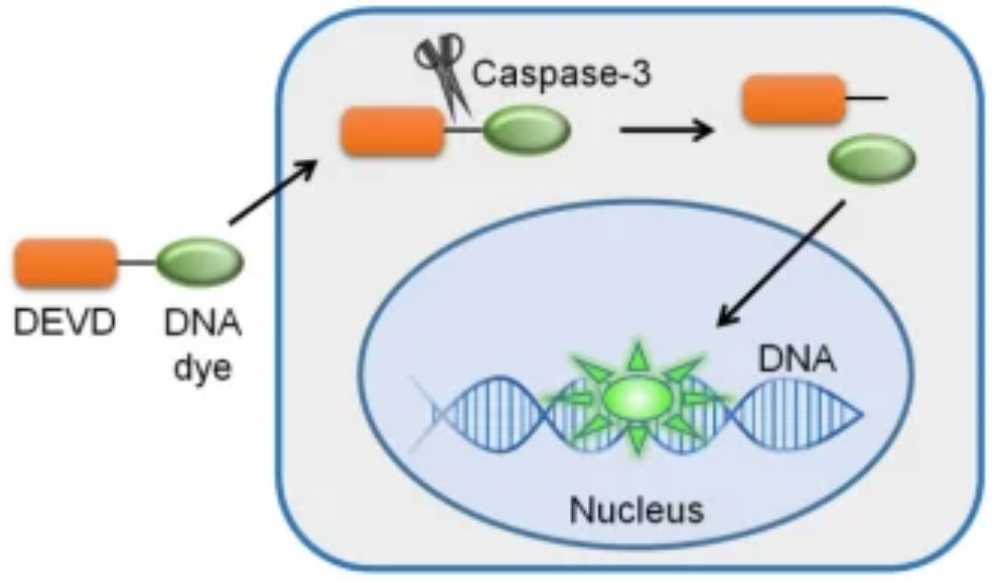
แคสเปส-3 เป็นตัวการสำคัญในกระบวนการอะพอพโทซิส เมื่อถูกกระตุ้นแล้ว มันจะก่อให้เกิดปฏิกิริยาต่อเนื่องมากมายจนนำไปสู่การสิ้นสุดของเซลล์
หากงานวิจัยของคุณมุ่งเน้นไปที่สัญญาณการตายของเซลล์นอกเหนือจากสถานะของเซลล์ การตรวจสอบ Caspase-3 สามารถให้หลักฐานที่ชัดเจนยิ่งขึ้นได้
| หมายเลขแคตตาล็อก | ชื่อสินค้า |
|---|---|
| ซีเอ1060 | ชุดตรวจจับ Caspase-3 ในเซลล์มีชีวิต แบบเรืองแสงสีเขียว |
การทดสอบนี้เข้ากันได้ดีกับการทดสอบการย้อมสี Annexin V เมื่อใช้ร่วมกัน จะเชื่อมโยงการเปลี่ยนแปลงของเยื่อหุ้มเซลล์ในช่วงแรกกับการเปลี่ยนแปลงที่เกิดขึ้นในระยะหลังของกระบวนการทำงาน
การตรวจหาการแตกตัวของ DNA ด้วยวิธี TUNEL
การทดสอบ TUNEL มักใช้ตรวจหาการตัดของ DNA ที่เกี่ยวข้องกับอะพอพโทซิส ในกระบวนการอะพอพโทซิส การแยกตัวของ DNA จะเผยให้เห็นปลาย 3’-OH เอนไซม์สามารถติดแท็กปลายเหล่านี้ด้วยเบสเรืองแสงหรือเบสที่เชื่อมต่อกับไบโอตินได้

เนื่องจาก TUNEL ตรวจจับการแตกหักของ DNA จึงเหมาะสำหรับการพิสูจน์ภาวะอะพอพโทซิสในระยะสุดท้าย การตรวจสอบชิ้นเนื้อ การตรวจสอบด้วยกล้องจุลทรรศน์ และข้อมูลสำรองหลังการทดสอบการทำงาน
| หมายเลขแคตตาล็อก | ชื่อสินค้า |
|---|---|
| T2130 | ชุดตรวจวิเคราะห์การตายของเซลล์แบบ TUNEL จาก Cell Meter สีเขียว |
| ที2193 | ชุดตรวจ SF640 TUNEL Apoptosis Kit, Far-Red |
| ที2194 | ชุดตรวจ SF555 TUNEL Apoptosis สีส้มแดง |
| ที2195 | ชุดตรวจ SF594 TUNEL Apoptosis สีแดง |
| ที2196 | ชุดตรวจ SF488 TUNEL Apoptosis สีเขียว |
| จี4890 | ชุดตรวจหาอะพอพโทซิส TUNEL สีเขียว |
| G4891 | ชุดตรวจหาภาวะอะพอพโทซิส TUNEL ด้วยวิธีโครโมเจนิค DAB |
| T2191 | ชุด Biotin Tunnel Apoptosis |
สำหรับรีวิวการทดสอบที่ครอบคลุมมากขึ้น คุณสามารถตรวจสอบได้ที่ หมวดหมู่ผลิตภัณฑ์ตรวจจับอะพอพโทซิส.
วิธีการทางสัณฐานวิทยาสำหรับการยืนยันด้วยสายตาอย่างรวดเร็ว
การระบายสีตามลักษณะทางกายภาพยังคงมีประโยชน์สำหรับการตรวจสอบอย่างรวดเร็ว วิธีการเหล่านี้ไม่สามารถใช้ทดแทนกันได้ การทดสอบระดับเส้นทาง. อย่างไรก็ตาม ข้อมูลเหล่านั้นก็ให้การสนับสนุนที่ชัดเจนต่อข้อค้นพบของคุณ
การย้อมสีเพื่อตรวจหาการควบแน่นของนิวเคลียสและความเสียหายของเยื่อหุ้มเซลล์
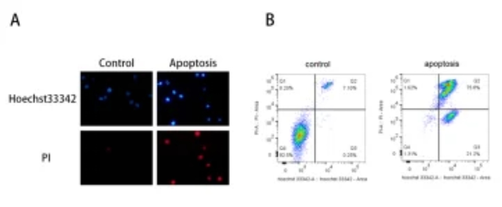
ในกระบวนการอะพอพโทซิส โครมาตินจะหดตัวแน่นขึ้น และนิวเคลียสอาจแตกออก สีเรืองแสงอย่างเช่น โฮเอชต์และพีไอ ช่วยให้คุณมองเห็นการเปลี่ยนแปลงเหล่านี้ได้อย่างชัดเจน
รูปแบบทั่วไปได้แก่:
เซลล์ปกติ: เรืองแสงสีแดงอ่อนและเรืองแสงสีน้ำเงินอ่อน
เซลล์ที่เกิดกระบวนการอะพอพโทซิส: เรืองแสงสีแดงอ่อนและเรืองแสงสีน้ำเงินเข้ม
เซลล์ที่ตายแล้ว: เรืองแสงสีแดงเข้มและเรืองแสงสีน้ำเงินเข้ม
| หมายเลขแคตตาล็อก | ชื่อสินค้า |
|---|---|
| CA1120 | ชุดย้อมสีคู่ Hoechst 33342/PI |
| โอ1320 | ชุดตรวจจับการซึมผ่านของเยื่อหุ้มเซลล์ Oxazole Yellow/PI เพื่อตรวจหาภาวะอะพอพโทซิส |
| ซีเอ1511 | ชุดตรวจวิเคราะห์วัฏจักรเซลล์และอะพอพโทซิส StaBright™ |
| จี3680 | ชุดย้อมสีอะพอพโทซิส Hoechst 33258 |
| ซีเอ1142 | ชุดอุปกรณ์ย้อมสีเรืองแสง AO |
หากคุณต้องการหลักฐานภาพถ่ายสำหรับเอกสาร การบรรยาย หรือการสแกนเบื้องต้น เครื่องมือปรับสีเหล่านี้จะมีประโยชน์อย่างมาก
วิธีการเลือกเวิร์กโฟลว์การตรวจจับอะพอพโทซิสที่เหมาะสม
กระบวนการอะพอพโทซิสที่มีประสิทธิภาพสูงสุดนั้นขึ้นอยู่กับคำถามที่คุณต้องการหาคำตอบ
| เป้าหมายการวิจัยของคุณ | แนวทางที่แนะนำ |
|---|---|
| ตรวจสอบการเกิดอะพอพโทซิสในระยะเริ่มต้น | แอนเน็กซิน วี/พีไอ |
| ศึกษาการบาดเจ็บของไมโตคอนเดรีย | เจซี-1, เจซี-10, เอ็มพีทีพี |
| ติดตามความเครียดที่เกี่ยวข้องกับแคลเซียม | โฟลว์-3, โฟลว์-4, โฟลว์-8 |
| ยืนยันการเกิดอะพอพโทซิสในระยะดำเนินการ | การทดสอบแคสเปส-3 |
| ตรวจจับการแตกหักของดีเอ็นเอ | การทดสอบ TUNEL |
| เพิ่มหลักฐานเชิงภาพ | การย้อมสี Hoechst/PI |
คำแนะนำสุดท้ายเพื่อข้อมูลอะพอพโทซิสที่ดีขึ้น
หากผลการศึกษาเกี่ยวกับอะพอพโทซิสดูไม่สม่ำเสมอ ปัญหาอาจไม่ได้เกิดจากหลักวิทยาศาสตร์เพียงอย่างเดียว แต่อาจเกิดจากการเลือกเทคนิคที่ไม่เหมาะสมกับขั้นตอนการทดสอบของคุณ
แนวทางที่มั่นคงกว่าคือการระบุว่าคุณต้องการหลักฐานสำหรับการเกิดอะพอพโทซิสในระยะเริ่มต้น ระยะกลาง หรือระยะสุดท้าย จากนั้นเลือกการทดสอบหลักหนึ่งอย่างและการทดสอบสำรองอีกหนึ่งอย่าง ต่อไป ปรับเทคนิคให้เข้ากับการตั้งค่าของคุณ สุดท้าย เชื่อมโยงผลการอ่านของคุณกับชีววิทยาที่การทดสอบแต่ละอย่างวัดได้อย่างแม่นยำ
เมื่อกระบวนการของคุณสอดคล้องกับคำถามในการศึกษา ผลลัพธ์ที่ได้จะเข้าใจง่ายขึ้น และมีคุณค่ามากขึ้นสำหรับการตัดสินใจ
ข้อสรุป
การตรวจจับอะพอพโทซิสจะง่ายขึ้นมากเมื่อคุณใช้การทดสอบร่วมกับปรากฏการณ์ทางชีวภาพที่คุณต้องการวัด การทดสอบศักยภาพของเยื่อหุ้มไมโทคอนเดรียช่วยในการตรวจจับสัญญาณความเครียดในระยะเริ่มต้น วิธีการที่ใช้แอนเน็กซิน V จำแนกขั้นตอนของอะพอพโทซิส การทดสอบแคสเปส-3 ตรวจสอบย้อนกลับกระบวนการ การทดสอบ TUNEL ตรวจสอบการแตกหักของ DNA
เพื่อให้ได้ผลลัพธ์ที่แม่นยำและมั่นคงยิ่งขึ้น ควรหลีกเลี่ยงการพึ่งพาเพียงสัญญาณเดียว แผนการวิเคราะห์แบบผสมผสานจะช่วยให้เห็นภาพรวมของการตายของเซลล์ได้ดียิ่งขึ้น และยังช่วยลดโอกาสในการตัดสินผิดพลาดจากสัญญาณเพียงอย่างเดียวอีกด้วย
ในการเตรียมการทดสอบอะพอพโทซิสที่จะเกิดขึ้น ให้เริ่มต้นด้วยเป้าหมายการศึกษาของคุณ เลือกการทดสอบหลักหนึ่งอย่าง และเพิ่มวิธีการตรวจสอบที่ตรงกันอีกหนึ่งวิธี เลือกชุดอุปกรณ์ ซึ่งเหมาะกับอุปกรณ์และวัสดุของคุณมากที่สุด กระบวนการนี้สามารถให้ข้อมูลที่คมชัดและน่าเชื่อถือยิ่งขึ้น นอกจากนี้ยังช่วยเพิ่มความเร็วในการศึกษาของคุณด้วย
เพื่อเปลี่ยนจากแนวคิดไปสู่การปฏิบัติ ให้พิจารณาผลิตภัณฑ์ทั้งหมด จับคู่เป้าหมายการตรวจจับของคุณกับตัวเลือกการทดสอบที่ระบุไว้ เลือกวิธีการที่ช่วยให้การทดสอบของคุณประสบความสำเร็จมากที่สุด
คำถามที่พบบ่อย
คำถามที่ 1: วิธีการตรวจจับอะพอพโทซิสแบบใดที่มีประโยชน์มากที่สุดสำหรับการวิเคราะห์ในระยะเริ่มต้น?
A1: การย้อมสี Annexin V/PI ถูกนำมาใช้กันอย่างแพร่หลายในการตรวจจับภาวะอะพอพโทซิสระยะเริ่มต้น เนื่องจากสามารถแยกแยะเซลล์ที่มีชีวิต เซลล์ที่อยู่ในระยะอะพอพโทซิสระยะเริ่มต้น และเซลล์ที่อยู่ในระยะอะพอพโทซิสระยะสุดท้ายได้
คำถามที่ 2: การสูญเสียศักยภาพของเยื่อหุ้มไมโทคอนเดรียเพียงอย่างเดียวสามารถพิสูจน์การเกิดอะพอพโทซิสได้หรือไม่?
A2: ไม่ใช่ค่ะ การตรวจนี้เป็นตัวบ่งชี้เบื้องต้นที่สำคัญ แต่ควรใช้ควบคู่กับการตรวจวิเคราะห์อื่น ๆ เช่น การย้อมสี Annexin V, การตรวจหา Caspase-3 หรือ TUNEL จะดีที่สุดค่ะ
คำถามที่ 3: ความแตกต่างหลักระหว่างการทดสอบ Caspase-3 และการทดสอบ TUNEL คืออะไร?
A3: การทดสอบ Caspase-3 ตรวจจับการกระตุ้นของกระบวนการอะพอพโทซิส ในขณะที่การทดสอบ TUNEL ตรวจจับการแตกตัวของ DNA ซึ่งมักปรากฏขึ้นในขั้นตอนต่อมา
คำถามที่ 4: การทดสอบแบบใดดีกว่าสำหรับการวิเคราะห์ด้วยเครื่องฟลูออโรไซโตเมตรี?
A4: การย้อมสี Annexin V/PI เป็นหนึ่งในตัวเลือกที่นิยมใช้มากที่สุดสำหรับการวิเคราะห์อะพอพโทซิสโดยใช้โฟลว์ไซโตเมทรี นอกจากนี้ โพรบแคลเซียมและสีย้อมศักยภาพไมโทคอนเดรียบางชนิดก็สามารถใช้สนับสนุนกระบวนการทำงานแบบโฟลว์ได้เช่นกัน
Q5: วิธีใดดีกว่าสำหรับการใช้กล้องจุลทรรศน์ฟลูออเรสเซนต์?
A5: การย้อมสี Hoechst/PI, การทดสอบ JC-1 และชุดตรวจ TUNEL แบบเรืองแสง ล้วนเป็นตัวเลือกที่เหมาะสม ขึ้นอยู่กับว่าคุณต้องการตรวจสอบลักษณะทางสัณฐานวิทยา ความเสียหายของไมโทคอนเดรีย หรือการแตกตัวของ DNA
Q6: ฉันจะแยกความแตกต่างระหว่างอะพอพโทซิสและเนโครซิสได้อย่างชัดเจนยิ่งขึ้นได้อย่างไร?
A6: วิธีการย้อมสีแบบคู่ เช่น Annexin V/PI หรือ Hoechst/PI สามารถช่วยแยกเซลล์ที่เกิดภาวะอะพอพโทซิสโดยที่เยื่อหุ้มเซลล์ยังคงสภาพสมบูรณ์ออกจากเซลล์ที่มีความเสียหายของเยื่อหุ้มเซลล์อย่างรุนแรงได้
Q7: วิธีที่ดีที่สุดในการทำให้ข้อมูลเกี่ยวกับการเกิดอะพอพโทซิสมีความน่าเชื่อถือมากขึ้นคืออะไร?
A7: ควรใช้การทดสอบอย่างน้อยสองวิธีที่เสริมกัน ตัวอย่างเช่น ใช้ Annexin V/PI สำหรับการเกิดอะพอพโทซิสในระยะเริ่มต้น และใช้ TUNEL หรือ Caspase-3 สำหรับการยืนยันผล
 ปรึกษา
 Whatsapp
 ทิ้งข้อความ
 ด้านบน