



ข่าวที่เกี่ยวข้อง
คำอธิบายเกี่ยวกับ TGF-β1: การกระตุ้น การส่งสัญญาณผ่าน SMAD บทบาทในโรคต่างๆ และการตรวจหาด้วยวิธี ELISA
ตารางเนื้อหา
TGF-β1 เป็นไซโตไคน์ที่ได้รับการศึกษาอย่างกว้างขวางและปรากฏในบริบทการวิจัยหลายด้าน การศึกษาบางเรื่องมุ่งเน้นไปที่การสร้างแผลเป็นและการซ่อมแซมเนื้อเยื่อ ในขณะที่การศึกษาอื่นๆ มุ่งเน้นไปที่การหลีกเลี่ยงภูมิคุ้มกันของเนื้องอก ภาวะพังผืดในไต การปรับโครงสร้างหัวใจ หรือกระบวนการที่เกี่ยวข้อง ดังนั้น สารวิเคราะห์ชนิดเดียวกันจึงมีความหมายที่แตกต่างกันไปขึ้นอยู่กับคำถามทางชีววิทยา
เหตุใด TGF-β1 จึงน่าสนใจ แต่ในขณะเดียวกันก็ศึกษาได้ยาก? มันสามารถยับยั้งการเพิ่มจำนวนเซลล์ในบางบริบทของเซลล์ ในขณะเดียวกันก็ส่งเสริมการซ่อมแซมเนื้อเยื่อ การเปลี่ยนแปลงจากเยื่อบุผิวเป็นเนื้อเยื่อเกี่ยวพัน การบุกรุก หรือการหลีกเลี่ยงภูมิคุ้มกันในบริบทอื่นๆ การส่งสัญญาณ TGF-β1 ที่ต่อเนื่องหรือผิดปกติยังสามารถกระตุ้นให้เกิดพังผืดได้ ดังนั้น การทดลองที่ให้ข้อมูลที่เป็นประโยชน์จึงควรพิจารณาถึงชนิดของเซลล์ ระยะของโรค กิจกรรมของตัวรับ การส่งสัญญาณ SMAD ในขั้นตอนถัดไป และรูปแบบโมเลกุลของ TGF-β1 ที่กำลังวัดอยู่
โซลาร์บิโอ จัดจำหน่ายแอนติบอดี ชุดตรวจ ELISA สารเคมีชีวภาพ ผลิตภัณฑ์ชีววิทยาของเซลล์ และเครื่องมือวิจัยที่เกี่ยวข้องสำหรับการศึกษาเกี่ยวกับกระบวนการเหล่านี้ นักวิจัยที่วางแผนขั้นตอนการทำงานที่กว้างขึ้นสามารถตรวจสอบรายการนี้ได้เช่นกัน โซลูชันการวิจัยด้านวิทยาศาสตร์ชีวภาพ ก่อนที่จะเลือกการทดสอบแต่ละรายการ
TGF-β1 คืออะไร?
สมาชิกสำคัญของกลุ่มซูเปอร์แฟมิลี TGF-β
ทรานส์ฟอร์มมิงโกรทแฟคเตอร์เบตา (TGF-β) หมายถึงกลุ่มของโพลีเปปไทด์ที่เป็นปัจจัยการเจริญเติบโตที่หลั่งออกมา ซึ่งอยู่ในกลุ่มซูเปอร์แฟมิลี TGF-β ที่ใหญ่กว่า สัตว์เลี้ยงลูกด้วยนมมี 33 ยีนที่เข้ารหัสลิแกนด์ของซูเปอร์แฟมิลีนี้ รวมถึงไอโซฟอร์ม TGF-β ทั้งสามชนิด (TGF-β1, TGF-β2 และ TGF-β3), แอคติวิน, โปรตีนโนดัล, ฮอร์โมนแอนติ-มุลเลเรียน, โปรตีนสร้างกระดูก และปัจจัยการเจริญเติบโตและการเปลี่ยนแปลงรูปร่าง
ลิแกนด์เหล่านี้มีโครงสร้างและหลักการส่งสัญญาณที่คล้ายคลึงกัน แต่แตกต่างกันในการใช้ตัวรับ เส้นทาง SMAD ระดับล่าง และผลกระทบทางชีวภาพ TGF-β1 เป็นไอโซฟอร์มที่ได้รับการศึกษามากที่สุด มีการแสดงออกอย่างกว้างขวางและควบคุมการเพิ่มจำนวนเซลล์ การแบ่งแยกเซลล์ การเคลื่อนย้ายเซลล์ การตายของเซลล์ การซ่อมแซมเนื้อเยื่อ และการตอบสนองทางภูมิคุ้มกัน
TGF-β1 มีความเกี่ยวข้องอย่างใกล้ชิดกับภาวะพังผืด มันถูกผลิตขึ้นโดยเซลล์หลายชนิด รวมถึงเซลล์ไฟโบรบลาสต์ เซลล์ภูมิคุ้มกัน และเซลล์เยื่อบุผิว การสมานแผลตามปกติจำเป็นต้องมีการลดทอนสัญญาณของ TGF-β การกระตุ้นอย่างต่อเนื่องอาจนำไปสู่การสะสมของเมทริกซ์นอกเซลล์มากเกินไปและทำให้เกิดภาวะพังผืดได้
โปรตีนที่มีการอนุรักษ์สูงในสิ่งมีชีวิตต่างชนิดกัน
ยีน TGFB1 ของมนุษย์ตั้งอยู่บนโครโมโซม 19q13.2 และเข้ารหัสโปรตีนก่อนการสังเคราะห์ (preproprotein) ซึ่งจะถูกประมวลผลเป็น LAP และเปปไทด์ TGF-β1 ที่ปลาย C-terminus ที่สมบูรณ์ TGF-β1 มีการอนุรักษ์ไว้อย่างสูงในสัตว์เลี้ยงลูกด้วยนม และแบบจำลองหนูถูกนำมาใช้กันอย่างแพร่หลายในการศึกษาหน้าที่ทางชีวภาพของมัน
การอนุรักษ์ลำดับสูงไม่ได้หมายความว่าจะขจัดความแตกต่างเฉพาะสายพันธุ์ การแสดงออกของตัวรับ ภูมิคุ้มกัน สภาพของเนื้อเยื่อ และแบบจำลองของโรค ล้วนสามารถส่งผลต่อผลลัพธ์ของการทดลองได้ ดังนั้น ผลการศึกษาจากแบบจำลองโรคไฟโบรซิสในหนูอาจให้ข้อมูลที่เป็นประโยชน์ แต่ไม่จำเป็นต้องนำไปใช้กับโรคไฟโบรซิสในมนุษย์โดยตรงเสมอไป
ความแตกต่างนี้มีความสำคัญเช่นกันเมื่อเลือกผลิตภัณฑ์ตรวจวัด ควรเลือกให้ตรงกับทั้งชนิดของสัตว์และเมทริกซ์ของตัวอย่างที่จะใช้กับการทดสอบที่ได้รับการรับรองแล้ว อย่าคิดว่าชุดทดสอบที่พัฒนาขึ้นสำหรับสัตว์ชนิดหนึ่งจะเหมาะสมกับสัตว์อีกชนิดหนึ่ง ปฏิกิริยาข้ามสายพันธุ์นั้นขึ้นอยู่กับผลิตภัณฑ์แต่ละชนิดและต้องได้รับการยืนยันจากข้อมูลการตรวจสอบความถูกต้อง
TGF-β1 ถูกผลิตขึ้นในรูปที่ไม่ทำงานก่อน
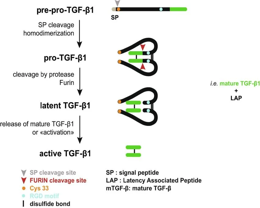
โดยทั่วไปเซลล์จะไม่หลั่ง TGF-β1 ออกมาในรูปของโมเลกุลส่งสัญญาณที่ออกฤทธิ์ได้ทันที แต่จะถูกสังเคราะห์ขึ้นในรูปของพรีโปรโปรตีนและถูกนำเข้าสู่เส้นทางการหลั่งโดยเปปไทด์ส่งสัญญาณที่ปลายด้าน N
ในออร์แกเนลล์กอลจิ สารตั้งต้นจะถูกตัดโดยเอนไซม์ฟูรินเพื่อสร้างเปปไทด์ที่เกี่ยวข้องกับภาวะแฝง (LAP) ที่ปลายด้าน N และเปปไทด์ TGF-β1 ที่ปลายด้าน C ที่สมบูรณ์ ไดเมอร์ของ LAP ยังคงจับกับไดเมอร์ของ TGF-β1 ที่สมบูรณ์โดยไม่ใช้พันธะโควาเลนต์ ทำให้คอมเพล็กซ์อยู่ในสภาวะแฝง
สารประกอบแฝงอาจถูกหลั่งออกมาหรือถูกกักเก็บไว้ที่ผิวเซลล์หรือในเมทริกซ์นอกเซลล์ผ่านทางพันธมิตรที่จับคู่กัน เช่น LTBP หรือ GARP ซึ่งเป็นการเพิ่มกลไกการควบคุมอีกชั้นหนึ่ง กล่าวคือ TGF-β1 สามารถอยู่ภายนอกเซลล์ได้โดยที่ไม่สามารถกระตุ้นตัวรับของมันได้
TGF-β1 ถูกกระตุ้นได้อย่างไร และ มันส่งสัญญาณอย่างไร?
การปลดปล่อยจากปมแฝง
TGF-β1 ที่อยู่ในสภาวะแฝงจะต้องถูกกระตุ้นก่อนที่การส่งสัญญาณไปยังตัวรับจะเริ่มต้นได้ อินทิกรินบางชนิดจะจับกับโมทีฟ RGD ใน LAP และออกแรงทางกล หรือทำงานร่วมกับโปรตีนอื่นๆ เพื่อเปิดเผยหรือปลดปล่อยไดเมอร์ TGF-β1 ที่สมบูรณ์แล้ว
เอนไซม์โปรตีเอสและการเปลี่ยนแปลงอื่นๆ ในสภาพแวดล้อมภายนอกเซลล์อาจส่งเสริมการกระตุ้นได้เช่นกัน กลไกหลักขึ้นอยู่กับเนื้อเยื่อและบริบทของโรค การกระตุ้นที่ควบคุมได้ช่วยสนับสนุนการซ่อมแซมบาดแผล ในขณะที่การกระตุ้นซ้ำๆ หรือต่อเนื่องสามารถรักษาการทำงานของไฟโบรบลาสต์และไมโอไฟโบรบลาสต์ในภาวะพังผืดเรื้อรังได้
ปริมาณ TGF-β1 ทั้งหมดไม่เท่ากับปริมาณ TGF-β1 ที่ออกฤทธิ์แล้ว ตัวอย่างอาจมี TGF-β1 ที่แฝงอยู่เป็นจำนวนมาก แต่มีลิแกนด์ที่ออกฤทธิ์อิสระเพียงเล็กน้อย ดังนั้น นักวิจัยจึงต้องพิจารณาว่าการทดสอบนั้นวัด TGF-β1 ที่ออกฤทธิ์ตามธรรมชาติ TGF-β1 ที่แฝงอยู่ หรือ TGF-β1 ทั้งหมดหลังจากขั้นตอนการกระตุ้นแล้ว
การจับกันของตัวรับเป็นจุดเริ่มต้นของสัญญาณ
TGF-β1 ที่ออกฤทธิ์จะจับกับตัวรับชนิดที่ 2 (TβRII) ก่อน จากนั้น TβRII ที่จับกับลิแกนด์แล้วจะดึงดูดตัวรับชนิดที่ 1 (TβRI/ALK5) เข้ามา ทำให้เกิดเป็นสารเชิงซ้อนส่งสัญญาณที่ผิวเซลล์
TβRII เป็นเอนไซม์ไคเนสชนิดเซริน/ทรีโอนีนที่ทำงานอย่างต่อเนื่อง ซึ่งทำหน้าที่ฟอสฟอริเลตและกระตุ้น TβRI ภายในคอมเพล็กซ์ตัวรับ-ลิแกนด์ ส่วน TβRIII หรือเบตาไกลแคน เป็นโครีเซปเตอร์ที่ไม่ใช่ไคเนส ซึ่งช่วยอำนวยความสะดวกในการนำเสนอลิแกนด์ไปยังคอมเพล็กซ์ตัวรับสัญญาณ โดยเฉพาะอย่างยิ่งสำหรับ TGF-β2
ปริมาณและการควบคุมของตัวรับก็มีความสำคัญเช่นกัน เซลล์สายพันธุ์ต่างๆ ที่สัมผัสกับความเข้มข้นของ TGF-β1 เท่ากัน อาจตอบสนองแตกต่างกันเนื่องจากมีการแสดงออกของระดับตัวรับที่แตกต่างกัน การกลายพันธุ์ของตัวรับ การนำเข้าสู่เซลล์ การแสดงออกของตัวรับร่วม และโปรตีนยับยั้ง สามารถเปลี่ยนแปลงความแรงของสัญญาณได้อีกด้วย
SMAD2 และ SMAD3 เป็นตัวกลางในการส่งสัญญาณแบบแคนอนิก
แบบคลาสสิก ทีจีเอฟ-เบตา/สมาด เส้นทางการส่งสัญญาณ กระบวนการนี้เกิดขึ้นโดยอาศัย SMAD2 และ SMAD3 เป็นหลัก เมื่อ TβRI ทำงาน มันจะทำการฟอสฟอริเลต SMAD ที่ถูกควบคุมโดยตัวรับเหล่านี้
TβRI ทำการฟอสโฟรีเลต SMAD2 และ SMAD3 ที่ถูกควบคุมโดยตัวรับ โปรตีนที่ถูกฟอสโฟรีเลตจะแยกตัวออกจากตัวรับและไปรวมตัวกับ SMAD4 ในไซโตพลาสซึม จากนั้นสารประกอบเหล่านี้จะสะสมอยู่ในนิวเคลียส ซึ่งพวกมันจะควบคุมการถอดรหัสยีนเป้าหมายร่วมกับพันธมิตรที่จับกับ DNA และตัวควบคุมร่วมในการถอดรหัส โปรแกรมการแสดงออกของยีนที่เกิดขึ้นจะขึ้นอยู่กับบริบทของเซลล์
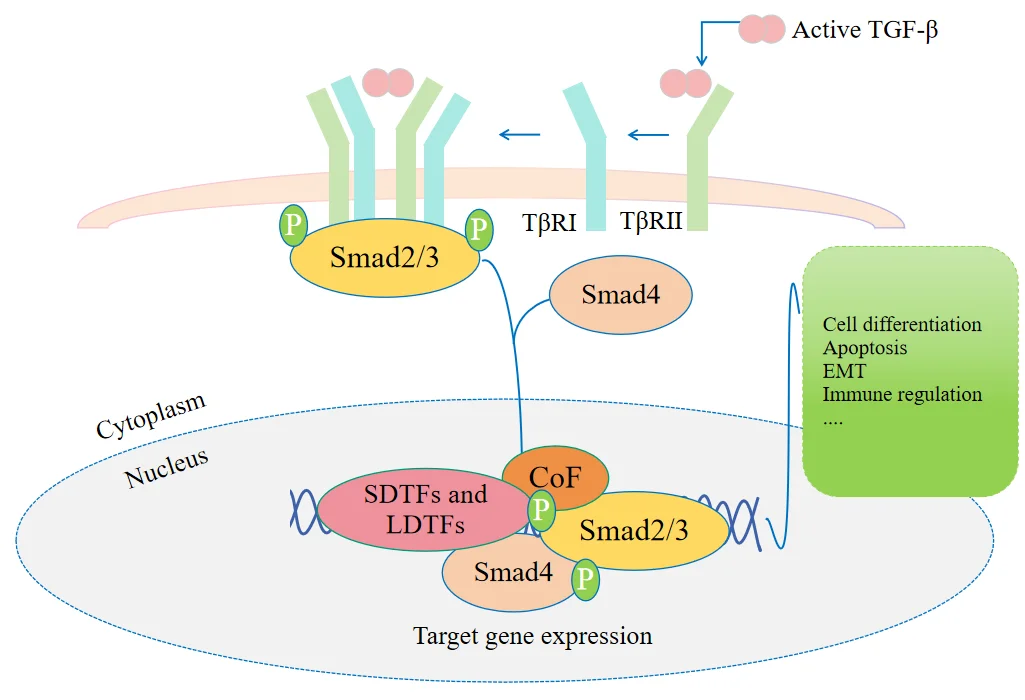
การส่งสัญญาณ TGF-β แบบดั้งเดิมสามารถส่งผลต่อการเปลี่ยนแปลงจากเซลล์เยื่อบุผิวเป็นเซลล์เนื้อเยื่อเกี่ยวพัน การตายของเซลล์ การควบคุมภูมิคุ้มกัน การสร้างเมทริกซ์นอกเซลล์ และการแบ่งแยกเซลล์ ผลลัพธ์ขึ้นอยู่กับชนิดของเซลล์ สภาวะการพัฒนาหรือโรค ระยะเวลาการส่งสัญญาณ และการโต้ตอบกับเส้นทางอื่นๆ การกระตุ้น SMAD ไม่ได้ก่อให้เกิดการตอบสนองที่ตายตัวเพียงอย่างเดียว
TGF-β1 มีบทบาทอย่างไรในภาวะสุขภาพและโรคต่างๆ?
การสมานแผลและพังผืด
TGF-β1 ส่งเสริมการดึงดูดและการกระตุ้นไฟโบรบลาสต์ และสามารถผลักดันให้เกิดการเปลี่ยนแปลงไปเป็นไมโอไฟโบรบลาสต์ได้ ไมโอไฟโบรบลาสต์ที่ถูกกระตุ้นจะผลิตคอลลาเจนและส่วนประกอบอื่นๆ ของเมทริกซ์นอกเซลล์ในปริมาณที่เพิ่มขึ้น ซึ่งช่วยในการซ่อมแซมเนื้อเยื่อ
ในกระบวนการสมานแผลตามปกติ ปฏิกิริยานี้จะลดลงเมื่อการซ่อมแซมเสร็จสมบูรณ์ ปัญหาจะเกิดขึ้นเมื่อการส่งสัญญาณ TGF-β ยังคงทำงานอย่างต่อเนื่อง คอลลาเจนจะสะสมมากขึ้น การสลายตัวของเมทริกซ์นอกเซลล์ลดลง และเนื้อเยื่อจะแข็งตัวขึ้นเรื่อยๆ
รูปแบบนี้เกิดขึ้นในภาวะพังผืดหลายชนิด ในแบบจำลองการทดลองของพังผืดในช่องท้อง TGF-β1 สามารถลดการแสดงออกของ E-cadherin ในขณะที่เพิ่ม α-SMA และคอลลาเจนชนิดที่ 1 ซึ่งสอดคล้องกับลักษณะฟีโนไทป์ที่สร้างเมทริกซ์แบบเมเซนไคม์มากขึ้น
การปรับโครงสร้างหัวใจ
TGF-β1 ยังได้รับการศึกษาในภาวะพังผืดในหัวใจและการปรับโครงสร้างของโพรงหัวใจด้วย ไฟโบรบลาสต์ที่ถูกกระตุ้นจะเพิ่มการผลิตคอลลาเจนชนิดที่ 1 และชนิดที่ 3 เมื่อเวลาผ่านไป การสะสมของเมทริกซ์มากเกินไปอาจลดความยืดหยุ่นของโพรงหัวใจและทำให้การทำงานของหัวใจบกพร่องได้
นักวิจัยมักตรวจสอบ TGF-β1 ร่วมกับการส่งสัญญาณของปัจจัยการเจริญเติบโตที่ได้จากเกล็ดเลือด, c-Abl, โปรตีนเมทริกซ์นอกเซลล์ และตัวบ่งชี้การอักเสบ การวัดปัจจัยใดปัจจัยหนึ่งเพียงอย่างเดียวอาจทำให้พลาดกระบวนการปรับเปลี่ยนโครงสร้างในวงกว้างได้
งานวิจัยที่เกี่ยวข้องกับการตั้งครรภ์
TGF-β1 ยังได้รับการศึกษาในงานวิจัยที่เกี่ยวข้องกับการตั้งครรภ์ด้วย เนื่องจากสัญญาณ TGF-β ควบคุมการสร้างรกและการบุกรุกของเซลล์โทรโฟบลาสต์ สัญญาณที่ผิดปกติมีความเกี่ยวข้องกับภาวะครรภ์เป็นพิษ ในแบบจำลองรกของมนุษย์ในการทดลอง มีรายงานว่า TGF-β1 กระตุ้นการผลิตอนุมูลอิสระที่ขึ้นอยู่กับ NADPH oxidase ผลการค้นพบนี้ควรได้รับการตีความในบริบทของแบบจำลองเฉพาะนั้นๆ มากกว่าที่จะมองว่าเป็นกลไกสากล
มะเร็งและการควบคุมภูมิคุ้มกัน
TGF-β1 มีบทบาทที่ซับซ้อนและขึ้นอยู่กับระยะของโรคมะเร็ง ในช่วงเริ่มต้นของการพัฒนาเนื้องอก เส้นทาง TGF-β สามารถทำหน้าที่เป็นตัวยับยั้งเนื้องอกโดยการยับยั้งการเพิ่มจำนวนและกระตุ้นให้เกิดอะพอพโทซิส ซึ่งทำหน้าที่เสมือนตัวเบรกการเติบโตของเนื้องอก
ในระยะหลังๆ เนื้องอกหลายชนิดสามารถหลีกเลี่ยงการยับยั้งการเจริญเติบโตที่เกิดจาก TGF-β ได้ ในบริบทนั้น การส่งสัญญาณ TGF-β ที่ต่อเนื่องหรือสูงขึ้นสามารถส่งเสริมการเปลี่ยนแปลงจากเซลล์เยื่อบุผิวเป็นเซลล์เนื้อเยื่อเกี่ยวพัน การรุกราน การแพร่กระจาย และการหลีกเลี่ยงระบบภูมิคุ้มกันได้
พฤติกรรมที่ขึ้นอยู่กับระยะของโรคนี้มีความสำคัญ การอธิบาย TGF-β1 ว่าเป็นเพียงตัวยับยั้งเนื้องอกหรือเพียงตัวส่งเสริมเนื้องอกนั้นง่ายเกินไป ผลลัพธ์ขึ้นอยู่กับชนิดของเนื้องอก พื้นฐานทางพันธุกรรม การทำงานของตัวรับ และระยะของโรค นักวิจัยสามารถติดตามการศึกษาเส้นทางใหม่ๆ ผ่านทาง Solarbio ได้ ข่าวสารด้านการวิจัยและการอัปเดตแอปพลิเคชัน.
นักวิจัยควรวัดระดับ TGF-β1 อย่างไร?
เริ่มต้นด้วยตัวอย่างและสายพันธุ์
คำถามแรกไม่ใช่ว่าชุดทดสอบใดมีขั้นตอนที่สั้นที่สุด แต่เป็นตัวอย่างประเภทใดที่จะนำมาทดสอบ เซรั่ม พลาสมา และสารละลายจากเซลล์เพาะเลี้ยงต้องการการจัดการที่แตกต่างกัน เมทริกซ์อื่นๆ รวมถึงเนื้อเยื่อที่บดละเอียดหรือสารละลายจากเซลล์ ควรใช้หลังจากตรวจสอบความถูกต้องของวิธีการทดสอบเฉพาะแล้วเท่านั้น เช่น การได้ผลลัพธ์ที่ดี ความเป็นเส้นตรงของการเจือจาง และผลกระทบของเมทริกซ์
การปลดปล่อยสารจากเกล็ดเลือดระหว่างการแข็งตัวของเลือดอาจทำให้ระดับ TGF-β1 ในซีรั่มที่วัดได้เพิ่มขึ้นอย่างมาก การเลือกใช้สารกันเลือดแข็ง ความล่าช้าในการประมวลผล สภาพการปั่นเหวี่ยง และการแช่แข็งและละลายซ้ำหลายรอบก็อาจส่งผลต่อผลลัพธ์ได้เช่นกัน ควรใช้โปรโตคอลการเก็บและประมวลผลที่กำหนดไว้เหมือนกันสำหรับทุกกลุ่ม และไม่ควรเปรียบเทียบซีรั่มกับพลาสมาโดยตรง
การเลือกสายพันธุ์ก็มีความสำคัญเช่นกัน Solarbio มีชุดตรวจ TGF-β1 ELISA สำหรับตัวอย่างจากหนู มนุษย์ หนูแรต หมู กระต่าย สุนัข ไก่ วัว และลิง
แยกระดับโปรตีนออกจากกิจกรรมของวิถีการทำงาน
ความเข้มข้นของ TGF-β1 ที่วัดได้สูงขึ้นไม่ได้หมายความว่าการส่งสัญญาณในขั้นตอนถัดไปจะแข็งแกร่งขึ้นเสมอไป โปรตีนส่วนใหญ่อาจยังคงอยู่ในสภาวะแฝง การแสดงออกของตัวรับอาจมีจำกัด หรือการส่งสัญญาณในขั้นตอนถัดไปอาจถูกยับยั้ง
กระบวนการทำงานที่ดีกว่าคือการรวมการวัดปริมาณ TGF-β1 เข้ากับการอ่านค่าเส้นทางต่างๆ เช่น SMAD2 และ SMAD3 ที่ถูกฟอสโฟรีเลต การแปลตำแหน่งของคอมเพล็กซ์ SMAD ในนิวเคลียส α-SMA คอลลาเจนชนิดที่ 1 E-cadherin หรือยีนเป้าหมายที่เลือกไว้
การเลือกใช้ชุดตัวบ่งชี้ที่เหมาะสมนั้นขึ้นอยู่กับคำถามวิจัย การศึกษาเกี่ยวกับพังผืดจะไม่ใช้ชุดตัวบ่งชี้เดียวกันกับโครงการวิจัยเกี่ยวกับภูมิคุ้มกันต่อเนื้องอก และการเพิ่มตัวบ่งชี้มากขึ้นก็ไม่ได้หมายความว่าจะดีกว่าเสมอไป การวัดผลแต่ละครั้งควรทดสอบขั้นตอนที่กำหนดไว้ในกลไกที่เสนอไว้
ใช้ชุดตรวจ ELISA ที่ตรงกับสายพันธุ์
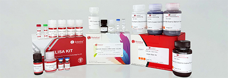
สำหรับตัวอย่างหนูนั้น ชุดตรวจ ElaBoX™ Mouse TGF-β1 ELISA มีตัวเลือกที่เหมาะสมกับสายพันธุ์สำหรับการตรวจวัดเชิงปริมาณในตัวอย่างที่ได้รับการตรวจสอบแล้ว โครงการวิจัยเกี่ยวกับมนุษย์สามารถใช้ตัวเลือกนี้ได้ ชุดตรวจ ElaBoX™ Human TGF-β1 ELISAโดยมีผลิตภัณฑ์แยกต่างหากสำหรับตัวอย่างจากหนู หมู กระต่าย สุนัข ไก่ วัว และลิง
กลุ่มผลิตภัณฑ์ชุด ELISA TGF-β1 ELISA ที่กว้างขึ้นของ Solarbio ได้แก่ SEKM-0035, SEKH-0316, SEKR-0012SEKP-0011 SECRT-0401, SEKC-0049, SEKCN-0005, SEKB-0305 และ SEKMY-0019
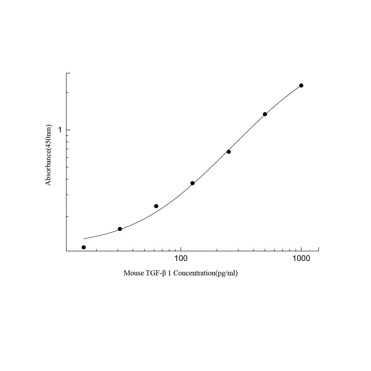
ก่อนทำการทดสอบตัวอย่างที่มีค่า โปรดตรวจสอบให้แน่ใจว่าการทดสอบรายงานค่า TGF-β1 ที่ออกฤทธิ์ตามธรรมชาติ หรือค่า TGF-β1 รวมที่กระตุ้นด้วยกรด สำหรับ SEKM-0035 นั้น เซรั่ม พลาสมา และสารละลายจากเซลล์เพาะเลี้ยงจำเป็นต้องกระตุ้นด้วยกรดก่อน จากนั้นจึงทำให้เป็นกลาง และต้องรวมปัจจัยการเจือจางที่ได้ลงในการคำนวณขั้นสุดท้ายด้วย นอกจากนี้ โปรดตรวจสอบประเภทตัวอย่างที่ได้รับการรับรอง ช่วงการตรวจจับ เส้นกราฟมาตรฐาน การฟื้นตัว ความแม่นยำ ความเป็นเส้นตรงของการเจือจาง และปฏิกิริยาข้ามสายพันธุ์ การทดสอบนำร่องระยะสั้นมักช่วยประหยัดเวลาได้มากกว่าการทำซ้ำแผ่นทดสอบเต็มรูปแบบในภายหลัง
Solarbio สามารถสนับสนุนเวิร์กโฟลว์ TGF-β1 ได้อย่างไร?
การคัดเลือกผลิตภัณฑ์โดยคำนึงถึงคำถามวิจัย
โครงการวิจัยเกี่ยวกับ TGF-β1 อาจต้องใช้ชุดตรวจ ELISA แต่ก็อาจต้องใช้แอนติบอดี สารเคมีสำหรับการเพาะเลี้ยงเซลล์ สารประกอบในกระบวนการสังเคราะห์โปรตีน การทดสอบทางชีวเคมี และเครื่องมือตรวจจับโปรตีนด้วย

การเลือกผลิตภัณฑ์ควรสอดคล้องกับคำถามทางชีววิทยา วัดปริมาณลิแกนด์เมื่อคำถามเกี่ยวข้องกับการหลั่งหรือปริมาณรวม ประเมินการฟอสโฟรีเลชันของตัวรับและ SMAD เมื่อเน้นที่การกระตุ้นวิถีการทำงาน เพิ่มตัวบ่งชี้พังผืด อะพอพโทซิส ความเครียดออกซิเดชัน หรือตัวบ่งชี้ภูมิคุ้มกันเมื่อประเมินการตอบสนองของเซลล์ในขั้นตอนถัดไป
แนวทางนี้ช่วยให้กระบวนการทำงานเชื่อมโยงกันอย่างเป็นระบบ และลดความเสี่ยงในการรวบรวมผลลัพธ์ที่ไม่เกี่ยวข้องกันซึ่งยากต่อการตีความร่วมกัน
การสนับสนุนทางเทคนิคสำหรับการวางแผนการทดสอบ
ประเภทของตัวอย่าง สายพันธุ์ ความเข้มข้นที่คาดหวัง และตารางการบำบัด ล้วนมีผลต่อแผนการทดสอบ Solarbio’s บริการทางเทคนิค สามารถช่วยเหลือนักวิจัยในการจับคู่ผลิตภัณฑ์กับขั้นตอนการทำงานที่วางแผนไว้ได้
การสนับสนุนทางเทคนิคมีประโยชน์อย่างยิ่งเมื่อต้องเปลี่ยนประเภทของตัวอย่างหรือเพิ่มตัวบ่งชี้ใหม่ โปรโตคอลที่ได้รับการตรวจสอบแล้วสำหรับซีรั่มอาจไม่สามารถนำไปใช้กับพลาสมา สารละลายจากการเพาะเลี้ยงเซลล์ หรือเมทริกซ์เนื้อเยื่อที่ยังไม่ได้รับการตรวจสอบได้โดยตรง
ก่อนขอรับการสนับสนุน โปรดเตรียมข้อมูลเกี่ยวกับสายพันธุ์ ตัวอย่าง รูปแบบของโรคหรือการรักษา ช่วงความเข้มข้นของ TGF-β1 ที่คาดหวัง ขั้นตอนการกระตุ้นที่วางแผนไว้ และผลลัพธ์ที่จะได้จากการวิเคราะห์
รักษาขั้นตอนการทำงานให้ง่ายต่อการทำซ้ำ
ผลการตรวจ TGF-β1 อาจเปลี่ยนแปลงได้เนื่องจากการจัดการตัวอย่าง การกระตุ้นด้วยกรดและการทำให้เป็นกลาง การเจือจาง ระยะเวลาการบ่ม หรือการล้างเพลท ความแตกต่างเล็กน้อยจะเห็นได้ชัดเจนมากขึ้นเมื่อค่าที่ได้อยู่ใกล้กับค่าต่ำสุดหรือสูงสุดของช่วงการทดสอบ
รักษามาตรฐานวิธีการเก็บรวบรวมและประมวลผลให้เหมือนกันในทุกกลุ่ม บันทึกสารกันเลือดแข็ง สภาวะการปั่นเหวี่ยง ระยะเวลาการเก็บรักษา ประวัติการแช่แข็งและการละลาย และปัจจัยการกระตุ้นและการเจือจาง ทดสอบสารมาตรฐานและสารควบคุมในทุกเพลท เมื่อเปรียบเทียบชุดตัวอย่างหรือการศึกษาต่างๆ ให้รักษาความสม่ำเสมอในการเตรียมตัวอย่างให้มากที่สุดเท่าที่จะเป็นไปได้
การควบคุมเหล่านี้เรียบง่าย แต่บ่อยครั้งที่ช่วยเพิ่มคุณภาพของข้อมูลได้มากกว่าการเพิ่มการทดสอบอีกครั้งในตอนท้าย
ข้อสรุป
TGF-β1 เป็นตัวควบคุมหลักของการซ่อมแซมเนื้อเยื่อ การเกิดพังผืด ความสมดุลของระบบภูมิคุ้มกัน การพัฒนา และชีววิทยาของมะเร็ง มันถูกผลิตขึ้นในรูปของสารประกอบแฝง และจะพร้อมใช้งานผ่านการกระตุ้นที่ถูกควบคุม และส่งสัญญาณเป็นหลักผ่านทาง TβRII, TβRI/ALK5, SMAD2, SMAD3 และ SMAD4
บทบาทของมันเปลี่ยนแปลงไปตามสภาพแวดล้อมทางชีววิทยา การส่งสัญญาณที่ควบคุมได้จะช่วยสนับสนุนการรักษาและการรักษาสมดุลของเนื้อเยื่อ ในขณะที่การส่งสัญญาณที่ต่อเนื่องหรือผิดปกติอาจกระตุ้นให้เกิดพังผืด การเปลี่ยนแปลงโครงสร้าง การรุกราน หรือการกดภูมิคุ้มกัน ดังนั้น การศึกษาที่ออกแบบมาอย่างดีจึงควรวัดมากกว่าแค่ความเข้มข้นของ TGF-β1 เพียงอย่างเดียว
ชุดตรวจ ELISA ที่ผ่านการตรวจสอบความถูกต้องของสายพันธุ์และเมทริกซ์ ตัวบ่งชี้ตัวรับและ SMAD และการทดสอบการทำงานขั้นปลายน้ำ ช่วยให้เห็นภาพเส้นทางได้ชัดเจนยิ่งขึ้น นักวิจัยที่วางแผนโครงการใหม่สามารถ ติดต่อทีมงาน Solarbio โดยระบุประเภทตัวอย่าง สายพันธุ์ และผลลัพธ์ที่คาดหวัง
เอฟเอคิว
คำถามที่ 1: TGF-β1 คืออะไร?
A1: TGF-β1 เป็นปัจจัยการเจริญเติบโตของโพลีเปปไทด์ที่หลั่งออกมาในกลุ่ม TGF-β ทำหน้าที่ควบคุมการเพิ่มจำนวนเซลล์ การแบ่งแยกเซลล์ การเคลื่อนย้ายเซลล์ การตายของเซลล์ การตอบสนองทางภูมิคุ้มกัน การซ่อมแซมเนื้อเยื่อ และการสร้างเมทริกซ์นอกเซลล์
คำถามที่ 2: TGF-β1 ถูกกระตุ้นได้อย่างไร?
A2: TGF-β1 ถูกสังเคราะห์ขึ้นในรูปของพรีโปรโปรตีน หลังจากถูกตัดโดยเอนไซม์ฟูรินแล้ว TGF-β1 ที่เจริญเต็มที่แล้วจะยังคงจับกับ LAP ในรูปแบบที่ไม่เกิดพันธะโควาเลนต์ในคอมเพล็กซ์ที่ไม่ทำงาน อินทิกริน โปรตีเอส และสัญญาณภายนอกเซลล์อื่นๆ สามารถเปิดเผยหรือปลดปล่อยลิแกนด์ที่ออกฤทธิ์แล้วได้
คำถามที่ 3: กลไกการส่งสัญญาณหลักของ TGF-β1 คืออะไร?
A3: TGF-β1 ที่ออกฤทธิ์จะจับกับ TβRII ซึ่งจะดึงดูดและฟอสโฟรีเลต TβRI/ALK5 TβRI ที่ถูกกระตุ้นจะฟอสโฟรีเลต SMAD2 และ SMAD3 จากนั้นโปรตีนเหล่านี้จะรวมตัวกับ SMAD4 สะสมอยู่ในนิวเคลียส และควบคุมการถอดรหัสยีนเป้าหมายร่วมกับพันธมิตรการถอดรหัสอื่นๆ
 ปรึกษา
 Whatsapp
 ทิ้งข้อความ
 ด้านบน