


Related news
Toluidine Blue O Staining Solution for Cells, Cartilage, Mast Cells, and Plant Tissue
Table of Contents
Toluidine Blue O is a familiar name in many staining rooms, but it should not be treated as one simple blue dye for every sample. A brain section, a cartilage section, a mast cell section, a fresh blood smear, and a plant stem section all have different staining needs. The target structure changes, the sample state changes, and the final color can change as well.
This is where product matching becomes important. Solarbio provides several Toluidine Blue O staining solutions for different samples, so researchers do not have to force one general formula into unrelated tissue types. For routine morphology work, this small choice often decides whether the result is clear enough to read under the microscope.
Why Toluidine Blue O Still Matters in Routine Staining
Sample matching is the first issue
Many staining problems do not come from the dye being useless. They come from using the wrong formula on the wrong sample. A product made for cartilage staining may not be the best choice for a fresh blood smear. A plant tissue staining solution should not be used in the same way as a mast cell stain.
Toluidine Blue O is useful because it can support several staining tasks, but the working solution must follow the sample. This is especially important for labs that handle animal tissue, plant tissue, and cell samples in the same project.
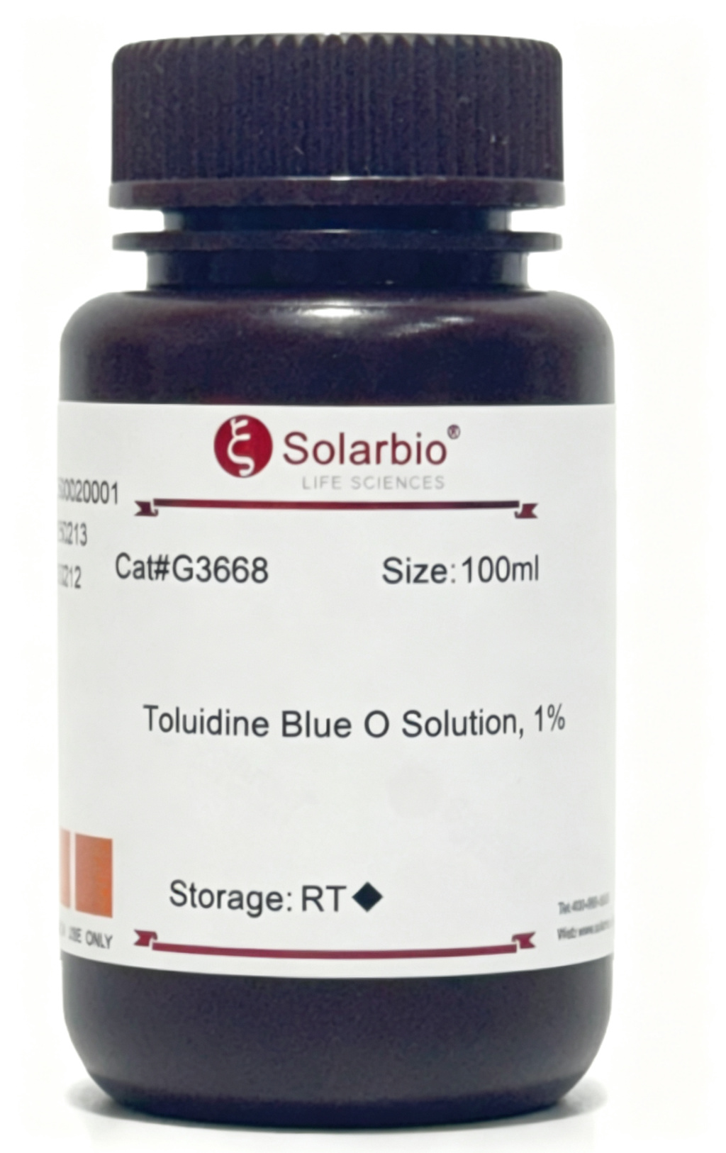
One dye can give more than one visual result
Toluidine Blue O can show blue, purple, reddish purple, or red tones. This is not a random color shift. It comes from the way the dye binds with different tissue components.
That color behavior makes it helpful in Nissl body staining, cartilage matrix staining, mast cell staining, plant tissue staining, and selected cell sample staining. The result is easy to observe when the sample preparation and staining time are controlled well.
The Chemistry Behind Toluidine Blue O Staining
A basic dye with clear chemical features
Toluidine Blue O is a thiazine dye. Its molecular formula is C14H12N2Cl2, its molecular weight is 293.21, and its CAS No. is 92-31-9. It belongs to the quinone-imine dye group and works as a basic dye.
Because the dye carries a positive charge, it has affinity for acidic tissue components. Its structure contains amino groups and quinone-type benzene ring chromophores, which help produce visible staining after binding with target structures.
Metachromasia is the key reason it is useful
The most practical feature of Toluidine Blue O is metachromasia. In regular staining, many structures appear blue. When the dye binds to certain acidic or sulfated substances, the color may shift toward purple, reddish purple, or red.
This explains why cartilage matrix and mast cell granules often show strong color contrast. Cartilage contains chondroitin sulfate and keratan sulfate. Mast cells contain heparin and histamine. These components respond well to basic dyes and create a clearer staining difference.
Researchers building a wider staining solution workflow can match sample preparation, staining product, and observation conditions before running larger batches.
Where Toluidine Blue O Works Best in Real Samples
Nervous tissue and Nissl bodies
In nervous tissue, Toluidine Blue O is often used for Nissl body staining. Nissl bodies are basophilic structures in neuronal cytoplasm. After staining, they may appear blue-purple, while neuronal nuclei often appear blue.
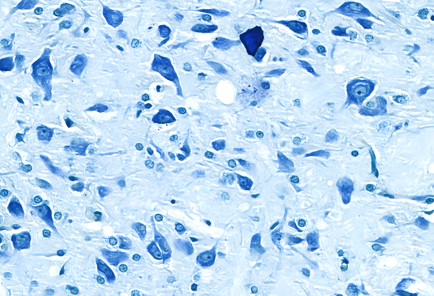
This type of staining is useful when researchers need to observe neuronal distribution and basic cell morphology in brain tissue sections. It gives a direct result without making the staining process too complicated.
Cartilage matrix and mast cells
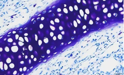
Cartilage matrix is one of the most common targets for Toluidine Blue O staining. Because the matrix contains acidic sulfated components, it can show a reddish purple metachromatic result. The background and nuclei usually remain blue, which gives useful contrast.
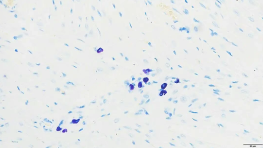
Mast cell staining follows a similar logic. Mast cell cytoplasm contains metachromatic substances such as heparin. With the right product, mast cell cytoplasm may appear purple-red, while nuclei appear blue. This helps researchers locate mast cells in tissue sections more easily.
Plant tissue and cell smears
Toluidine Blue O also works in plant tissue staining. It can help distinguish lignified cell walls from non-lignified areas. Lignified cell walls may show blue-green staining, while other cell wall areas may appear purple-blue.
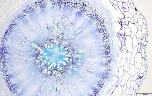
img.Toluidine Blue O plant tissue staining showing lignified and non-lignified cell wall differences.webp
For cell samples, Toluidine Blue O can be used when the target cells contain sulfated glycosaminoglycans. Chondrocytes and basophilic cells may show stronger purple-red or red staining, while other cells remain blue.
Solarbio Toluidine Blue O Products and Application Choices
For Nissl and cartilage staining
G1436 Nissl Stain Solution(Toluidine Blue Method) is suitable for Nissl body staining. In a mouse brain coronal paraffin section, Nissl bodies can appear blue-purple, and neuronal nuclei appear blue.
G2543 Toluidine Blue Cartilage Stain Solution is designed for cartilage staining. In a mouse nasal bone paraffin section, cartilage can show reddish purple staining, while the background and nuclei appear blue.
For cells, mast cells, and plant sections
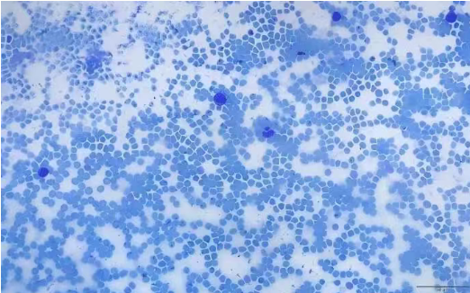
G3660 Toluidine Blue Stain Solution(For Cells) is made for cell sample staining. In a fresh mouse whole blood smear, lymphocytes and basophilic cells may appear purple-red, while red blood cells appear blue.
G3670 Mast Cell Stain Solution(Toluidine Blue Method) is used for mast cell staining. In mouse heart and lung paraffin sections, mast cell cytoplasm may appear purple-red, and nuclei appear blue.
G4807 Plant Tissue Stain Solution(Toluidine Blue Method) is suitable for plant tissue sections. In a cabbage stem paraffin section, lignified cell walls can appear blue-green, while non-lignified cell walls show purple-blue staining.
Related Staining Reagents for Broader Lab Work
Toluidine Blue O is not always the only stain needed
Many labs keep several staining products on hand. Toluidine Blue O is useful for specific metachromatic staining needs, but HE staining, Alcian Blue staining, other Nissl staining methods, and mast cell staining kits are also common in tissue morphology workflows.
Related products include G1120 Hematoxylin-Eosin(HE) Stain Kit, G1121 Modified Hematoxylin-Eosin(HE) Stain Kit, G1126 Hematoxylin-Eosin(HE) Stain Kit III, G1430 Nissl Stain Kit(Cresyl Violet Method), G1434 Nissl Stain Kit(Methylene Blue Method), G1560 Alcian Blue Stain Kit, pH 2.5, G2541 Cartilage Stain Kit(Alcian Blue Method, pH 1.0), and G2542 Cartilage Stain Kit(Alcian Blue Method, pH 2.5).
A practical staining shelf saves time
For a lab that handles many tissue types, keeping only one stain is rarely enough. A better setup is to prepare products by application. Brain tissue, cartilage, mast cells, plant sections, and routine tissue morphology may each need different staining kits.
Researchers comparing staining choices across tissue types can check Solarbio’s research pathways page. New product updates and staining-related information can also be found through technical news.
Practical Notes Before Ordering Toluidine Blue O Stains
Start with the sample and target structure
Before ordering, confirm the sample type first. If the sample is nervous tissue, G1436 is the more direct match. If the sample is cartilage, G2543 should be considered first. If the work involves fresh cell smears, G3660 is more suitable. If the target is mast cells, G3670 is the focused option. If the sample is plant tissue, G4807 fits the application better.
The sample format also matters. Paraffin sections, frozen sections, fresh blood smears, and plant sections do not behave the same way during staining and washing. Product selection should follow both the biological target and the sample form.
Keep the method stable once the result works
For repeated projects, consistency is worth recording. Keep the same product code when possible. Record section thickness, fixation method, staining time, washing time, and microscope settings.
Small changes can affect the final color depth. This is especially true when the staining result is used for comparison across batches. Labs that need method support can check Solarbio’s service page. If the product choice is still unclear, it is better to contact Solarbio with the sample type, target structure, and staining purpose before placing an order.
Conclusion
Toluidine Blue O is useful because it is simple to use, but it is not a careless choice. The stain works best when the product formula matches the sample. Brain tissue, cartilage, mast cells, fresh cell smears, and plant tissue sections all need different attention.
Solarbio provides several Toluidine Blue O staining solutions, including G1436, G2543, G3660, G3670, and G4807. These products cover common staining needs from Nissl bodies to cartilage matrix, mast cells, cell samples, and plant cell walls. For labs that want cleaner staining and fewer repeated trials, choosing by sample type is the safer route.
FAQ
Q1: What is Toluidine Blue O mainly used for?
A1: Toluidine Blue O is mainly used for staining acidic tissue components. It is commonly used in Nissl body staining, cartilage matrix staining, mast cell staining, plant tissue staining, and some cell sample staining.
Q2: Is Toluidine Blue O only suitable for animal tissue sections?
A2: No. It can also be used for plant tissue sections. For plant samples, it can help distinguish lignified and non-lignified cell walls.
Q3: Which Solarbio product is suitable for Nissl body staining?
A3: G1436 Nissl Stain Solution(Toluidine Blue Method) is suitable for Nissl body staining. It can show Nissl bodies in blue-purple tones in nervous tissue sections.
Q4: Which product should be used for cartilage staining?
A4: G2543 Toluidine Blue Cartilage Stain Solution is designed for cartilage staining. It helps show cartilage matrix with reddish purple staining, while the background and nuclei appear blue.
Q5: Can Toluidine Blue O be used for mast cell staining?
A5: Yes. G3670 Mast Cell Stain Solution(Toluidine Blue Method) is suitable for mast cell staining. Mast cell cytoplasm can show purple-red staining because of metachromasia.
Q6: Why do different samples show different colors with Toluidine Blue O?
A6: Different colors come from the chemical components in the sample. Acidic and sulfated substances can cause metachromatic staining, while other structures may appear blue.
Q7: Can one Toluidine Blue O solution be used for every sample type?
A7: It is not recommended. Different sample types need different formulations. A cartilage stain may not be the best choice for fresh cell smears, mast cell sections, or plant tissue sections.
Q8: What should be checked before choosing a Toluidine Blue O product?
A8: The sample type, target structure, and sample format should be checked first. Section thickness, fixation method, staining time, and washing time should also be recorded for repeatable results.
 Consult
 Whatsapp
 Leave Message
 top
top