



Related news
Double Immunodiffusion Assay: Principle, Protocol, Results, and Troubleshooting
Table of Contents
The double immunodiffusion assay is a relatively simple immunological test. However, a few minor mistakes during the setup can leave you with a blank agarose plate even after hours of incubation. Inappropriate antibody concentration, the bad shape of the wells, the drying out of the gel during the incubation or simply the wrong time of inspection of the plate can cause these problems.
This method can be used for observing direct antigen-antibody precipitation. No label or instrument is required for the detection of the reaction. Soluble antigen and its corresponding antibody diffuse as a solution through an agarose gel towards each other. When they meet at a suitable concentration ratio, a visible white precipitin line forms.
For laboratories running routine immunology and protein work, the assay can be built with standard reagents available through the Solarbio product portfolio, including immune serum, purified proteins, agarose, saline, and related laboratory materials.
What Is a Double Immunodiffusion Assay?
The technique of Double immunodiffusion is based on the movement of soluble antigens and antibodies in a semisolid gel. Antigens are placed in one well of the gel and the corresponding antibodies in the other well. Both antigens and antibodies then diffuse out of their respective wells at the same time.
Where the two components reach a suitable proportion, antigen-antibody complexes become large enough to precipitate. This creates a white line that can usually be seen directly on the plate.
Why a Precipitin Line Forms
The visible line depends heavily on the balance between antigen and antibody. Too much of either component can move the reaction away from the concentration range where precipitation is easiest to observe.
This is also why the location of the line can change. When antigen and antibody concentrations are relatively similar, the precipitin line usually appears between the two wells. When their concentrations are different, the line tends to form closer to the well containing the lower-concentration component.
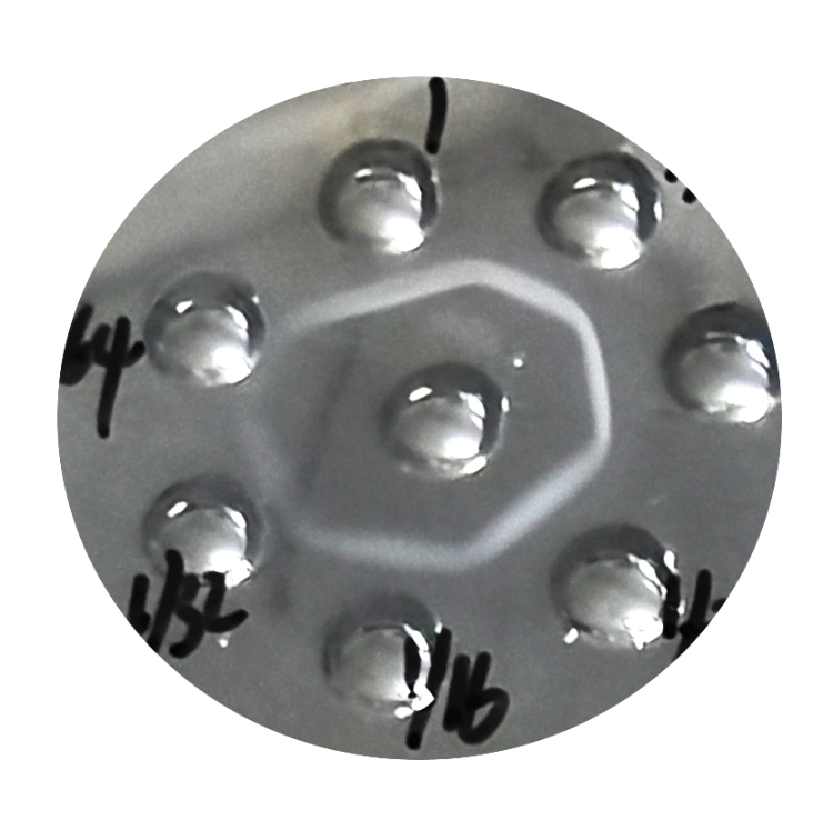
The basic reaction may look simple, but antibody quality and concentration still matter. Researchers who want a broader background on immunoglobulin structure and binding can also read What Biological Molecule Are Antibodies?.
What Can Double Immunodiffusion Be Used For?
The method can be used for qualitative identification of an antigen or antibody. It can also help with antibody titer assessment, purity analysis, and comparison of antigens or antibodies from different sources.
For this reason, double immunodiffusion remains useful in teaching laboratories and basic immunology work where a visible antigen-antibody reaction is enough to answer the experimental question.
It is different from quantitative methods such as ELISA. There is no enzyme-conjugated detection system and no absorbance reading. The main result is the formation, position, and visibility of the precipitin line.
Materials Needed for Double Immunodiffusion
A typical experiment starts with serum and corresponding antiserum, or a purified protein paired with an antibody that recognizes it. The gel system requires agarose together with physiological saline or another suitable buffer.
Petri dishes, pipettes, pipette tips, a well punch, and basic incubation equipment are also needed.
Choosing the Antigen and Antibody
The first thing to check is whether the antigen and antibody are actually specifically recognizes. A clean plate will not produce a useful line if the antibody does not recognize the antigen.
Concentration also deserves attention. For purified antibody, a concentration about 1 mg/mL as a starting reference is a practical starting point in this type of assay. If the sample is too dilute, the precipitate may be too weak to see even when binding is taking place.
Solarbio offers immune serum products for immunological work. One example is Goat anti-Human IgG (Immune serum) SA101, which can be paired with a suitable human IgG antigen according to the experimental design.
Preparing the Agarose Medium
A 1% agarose solution can be prepared in physiological saline. Phosphate buffer or barbiturate buffer may also be used depending on the laboratory method.
The agarose needs to dissolve fully. An uneven gel can change diffusion and make the final line harder to interpret. The melted agarose is generally cooled to around 50 to 60°C before it is poured into a Petri dish.
A gel thickness of about 3 to 5 mm works for the plate setup described here. Agarose A8201 is one of the supporting reagents that can be used when preparing an agarose-based immunodiffusion medium.
How to Perform the Double Immunodiffusion Assay
Good results depend more on careful plate preparation than on complicated equipment. The wells need to be neat, the gel must stay moist, and the samples should remain inside their assigned wells.
Preparing the Plate and Wells
After the agarose has completely set, wells can be punched into the gel. A common layout uses one central well surrounded by several outer wells.
A well diameter of around 3 mm can be used, with approximately 5 to 6 mm between the central well and the surrounding wells. Clean well edges matter because damaged agarose can cause uneven diffusion.
After the gel plugs have been removed, check the bottoms of the wells carefully. The sample should stay in the well rather than leaking underneath the agarose layer.
The plate can then be marked so that every surrounding well is easy to identify later. This small step becomes useful when several antigen concentrations or antibody dilutions are being compared on the same plate.
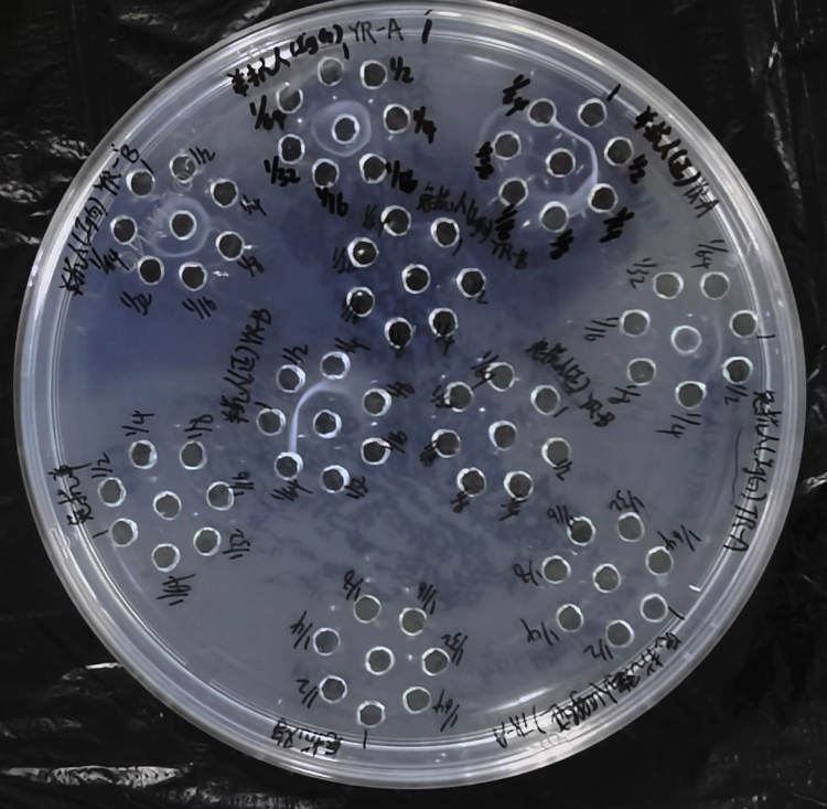
Loading Antigen and Antibody
Antigen and antibody are added separately to the prepared wells. When a dilution series is being tested, the different concentrations can be loaded into the surrounding wells in order. The undiluted sample can be placed in the central well, depending on the experimental arrangement.
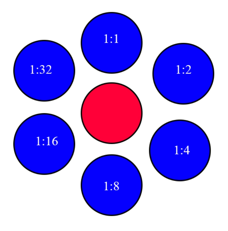
A physiological saline control can be included as a negative control.
Fill the wells carefully. Too little sample may produce a weak line, while overfilling can cause sample to spill onto the gel surface and interfere with the diffusion pattern.
Researchers working with several immunological reagents in the same project may also find the practical reagent selection discussion in Biochemical Reagents for Immunology & Protein Research useful when setting up the broader workflow.
Incubation and Observation
Once loading is complete, cover the plate and place it in a humid chamber. The gel should remain moist throughout incubation.
The plate can be incubated at 37°C and observed during a period of roughly 10 to 36 hours. During a first experiment, it is worth checking the plate several times rather than waiting until the end.
For product selection or matching antigens, immune sera, and supporting reagents to a research workflow, researchers can contact Solarbio with the sample type and planned assay conditions.
Why Is There No Precipitin Line?
A missing line does not automatically mean that the antibody failed. Double immunodiffusion depends on diffusion, concentration, gel condition, and observation time at the same time.
Check the Gel Before Changing the Antibody
Start with the agarose. It should be completely dissolved before the plate is poured. The surface should be even, and the wells should have clean edges.
The humid chamber also needs enough moisture. If the gel dries during incubation, normal antigen and antibody diffusion will be disturbed.
Well loading deserves another look. A partly filled well may simply contain too little antigen or antibody to form a clearly visible precipitate.
Solarbio provides broader immunology and life science research solutions for laboratories working across sample preparation, protein research, antibody assays, and related workflows.
Check Concentration and Observation Time
If the gel looks good, the next question is concentration. Very low antibody concentration is a common reason for weak or absent lines.
Antigen concentration can cause the same problem. When testing an unfamiliar sample, a dilution series is often more useful than relying on one concentration.
Timing can also catch people out. If the plate is checked too early, the antigen and antibody may not have diffused far enough to form a visible line. If it is left too long, the line may become difficult to see. Recording several time points gives a much clearer picture.
Reagents Commonly Used in the Assay
A basic double immunodiffusion experiment may use Bovine IgG Powder SP033 together with Rabbit anti-Bovine IgG SA233, or Human IgG Powder SP001 together with Goat anti-Human IgG SA101.
Blocking Normal Human Serum SL010 may be used in related immunological work, while Normal saline IN9004 and Agarose A8201 support preparation of the diffusion system.
These products serve different jobs. The IgG protein acts as the antigen in an appropriate setup, the immune serum supplies the corresponding antibody, and agarose provides the medium through which both components move.
For laboratories moving from a standard assay to custom proteins or antibodies, Solarbio also provides technical services covering antibody and protein-related research needs.
Conclusion
Double immunodiffusion does not need a complicated instrument, but it does need a controlled setup. The agarose has to be uniform, the wells need clean edges, the gel must stay moist, and antigen and antibody concentrations need to fall within a useful range.
When a plate shows no precipitin line, changing everything at once usually makes troubleshooting harder. Check the agarose first, then the wells and loading volume, followed by sample concentration and incubation time.
For product selection or questions about matching an antigen, immune serum, or supporting reagent to a research workflow, researchers can contact Solarbio with the sample type and planned assay conditions.
FAQ
Q1: What is the principle of a double immunodiffusion assay?
A1: Soluble antigen and antibody are placed in separate wells in an agarose gel. Both diffuse through the gel. When they meet at a suitable concentration ratio, antigen-antibody complexes precipitate and form a visible white line.
Q2: What agarose concentration is commonly used for double immunodiffusion?
A2: A 1% agarose solution can be used for the plate setup described here. The agarose should be fully dissolved before pouring so that diffusion takes place through a uniform medium.
Q3: Why is there no precipitin line in my immunodiffusion assay?
A3: Common causes include low antigen or antibody concentration, too little sample in the wells, poorly formed wells, uneven agarose, a dry plate, or unsuitable observation time. Each factor should be checked separately.
Q4: How concentrated should the antibody be?
A4: For purified antibody, a concentration around or above 1 mg/mL can be used as a practical reference for this assay. The suitable concentration may still vary with the antigen and antibody pair.
Q5: How long should a double immunodiffusion plate be incubated?
A5: The plate can be incubated at 37°C and observed during approximately 10 to 36 hours. During the first run, checking the plate every 5 to 10 hours can help identify when the precipitin line is most visible.
Q6: Why does the precipitin line form closer to one well?
A6: Line position is affected by antigen and antibody concentration. When the concentrations are unequal, the visible line generally appears closer to the well containing the lower-concentration component.
Q7: Can double immunodiffusion be used to measure antibody titer?
A7: Yes. The assay can be used for qualitative identification and titer assessment. It can also support purity analysis and comparison of antigen or antibody samples from different sources.
Q8: Is double immunodiffusion the same as ELISA?
A8: No. Both depend on specific antigen-antibody recognition, but double immunodiffusion produces a visible precipitin line in agarose. ELISA uses an enzyme-linked detection system and is commonly read through a measurable color signal.
 Consult
 Whatsapp
 Leave Message
 top