


Related news


EGFR Small-Molecule Inhibitors: What Actually Matters in Mechanism, Resistance, and Compound Selection
Table of Contents
EGFR is one of those targets that keeps coming back in cancer research. It appears in pathway diagrams, inhibitor screening, and almost every NSCLC-related project. But once the experiment moves beyond theory, the focus shifts quickly. The question is no longer what EGFR does, but which inhibitor to use, and whether the data really reflect pathway inhibition.
Different labs run into this at different stages. One group may only need to show reduced EGFR phosphorylation. Another may be working with a T790M-resistant model. Some focus on apoptosis, others on migration or long-term resistance. These are all EGFR-related questions, but the experimental setup behind them is not the same.
What are EGFR inhibitors?
EGFR belongs to the HER/ErbB receptor family and functions as a transmembrane tyrosine kinase. Under normal conditions, ligand binding triggers receptor dimerization and phosphorylation, which then activates downstream signaling.
Under pathological conditions, especially in various solid malignant tumors such as non-small cell lung cancer, colorectal cancer and head and neck squamous cell carcinoma, the EGFR gene frequently undergoes point mutations, deletion mutations, insertion mutations or overexpression. These alterations induce abnormal receptor structure, render it uncoupled from normal physiological regulation and keep it constitutively activated. This process further drives the unlimited proliferation, local invasion and distant metastasis of tumor cells, while inhibiting tumor cell apoptosis. Accordingly, EGFR acts as a core driver target for tumor initiation and progression.
This is why EGFR shows up so often in:
Non-small cell lung cancer
Colorectal cancer
Head and neck squamous cell carcinoma
How EGFR Inhibitors Work in Real Experiments
Most EGFR small-molecule inhibitors target the ATP-binding pocket of the kinase domain. By blocking ATP binding, they reduce phosphorylation and weaken downstream signaling.
On paper, this looks consistent. In practice, results vary a lot between models.
For example:
In an EGFR-mutant cell line, Gefitinib may reduce viability and trigger apoptosis within 48 hours
In another model with partial resistance, the same concentration may only affect phosphorylation
In a resistant cell line, the response may be minimal even at higher doses
These differences come from mutation background, pathway compensation, and experimental conditions. Even factors like cell density or how the stock solution was prepared can shift the outcome.
Because of this, compound selection should not rely only on reported IC50 values. A compound that works well in one paper may behave differently in another lab setup.
Key Pathways to Check, Not Just EGFR Itself
EGFR does not act alone. Once activated, it connects to several signaling pathways that control cell behavior.
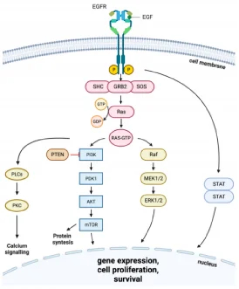
RAS/RAF/MAPK
Activated sequentially by EGFR, this pathway regulates downstream gene expression and drives cell cycle progression and proliferation. Phosphorylated ERK is commonly used to assess its activity in experiments.
PI3K/AKT
It transmits cell survival signals and inhibits apoptosis. Sustained activation of this pathway is a major cause of resistance to EGFR-targeted drugs and impaired cell death.
JAK/STAT3
It regulates both cell proliferation and immune responses, and plays a vital role in long-term treatment models, tumor microenvironment and drug resistance research. AG-490 can be applied to specifically block this pathway.
Not every experiment needs to cover all three pathways. But relying only on cell viability is rarely enough. Adding at least one downstream readout makes the data easier to interpret later.
Resistance Is Part of the Workflow
EGFR inhibitor studies almost always involve resistance at some point.
Two mutations come up repeatedly:
T790M: reduces binding of early-generation inhibitors.For T790M-focused resistance models, CO-1686 (Rociletinib) can be added as a targeted EGFR inhibitor option when comparing resistant-cell responses.
C797S: affects covalent binding of some irreversible inhibitors
Beyond mutation-driven resistance, EGFR blockade may be weakened by bypass signaling. In some models, MET or HER2 activation keeps downstream pathways active even when EGFR kinase activity is reduced. Other cells may not show strong molecular changes at the beginning, but later display a more invasive phenotype or a weaker treatment response. These patterns are easy to miss if the experiment only captures an early time point.
Weak inhibition does not always mean that the compound quality is poor. In T790M-positive models, for example, a general EGFR inhibitor may give limited results simply because it does not match the resistance background. The first step is therefore to align the inhibitor with the mutation profile and the purpose of the experiment.
Matching the Inhibitor to the Cell Model
Before selecting an EGFR inhibitor, check the points that most often change the experimental outcome:
Mutation status, including exon 19 deletion, L858R, T790M, and C797S
Whether the inhibitor is reversible or irreversible
Target range, such as EGFR-specific inhibition or broader HER family activity.Pelitinib may also be included in EGFR inhibitor comparison groups when the experiment needs another EGFR-focused compound for dose-response testing or phosphorylation readout validation.
Solubility and storage conditions
Planned assay endpoints
When the study design requires an irreversible EGFR-selective inhibitor, CL-387785 can be considered for comparing phosphorylation, viability, or downstream pathway responses with other EGFR inhibitor types.
For example:
Osimertinib is often used for T790M-related studies
Afatinib or Dacomitinib may be chosen for broader ErbB inhibition
Lapatinib or Neratinib are useful when HER2 signaling is involved
Solubility should be checked before the first treatment, not only after inconsistent data appear. Some compounds are listed as soluble in DMSO, yet may still precipitate during stock preparation or dilution into culture medium. Once precipitation occurs, the actual delivered dose can differ from the planned dose. This problem is easy to miss at the beginning, but it often becomes visible when replicate wells or repeat experiments start to separate.
Multidimensional Detection System for EGFR Research
A single assay rarely tells the full story in EGFR work.
Common endpoints include:
Cell viability
Apoptosis
Cell cycle distribution
EGFR phosphorylation
ERK or AKT phosphorylation
Migration and invasion
A practical workflow usually begins with a concentration gradient to define the response window. At this stage, the concentration gradient should be used to map the response pattern, not just to pick a dose with lower viability. A viability change may come from apoptosis, delayed proliferation, cell-cycle arrest, or non-specific cytotoxicity. For that reason, apoptosis assays or cell-cycle analysis are often needed after the first viability screen.
If the main question is pathway inhibition, p-EGFR should be measured together with downstream markers such as p-ERK or p-AKT. Comparing these results with the viability data helps separate a true signaling effect from a general loss of cell fitness. When the experiment depends on only one endpoint, the interpretation remains narrow.
Reagents Matter More Than Expected
In EGFR studies, the inhibitor is only one part of the system. Buffers, detection reagents, and assay kits all contribute to the final result.
Common issues seen in routine experiments include:
Buffer conditions affecting enzyme activity
Variability between reagent batches
Impurities causing off-target effects
Reliable reagent systems usually maintain tight quality control and low batch variation, which helps keep results consistent .
For labs running repeated experiments or comparing data across projects, this consistency becomes more important than the initial cost.
A Note on Sourcing Compounds and Supporting Reagents
EGFR inhibitor experiments usually require several types of supporting reagents. The same treatment group may be analyzed by viability assays, apoptosis detection, western blotting, and cell culture-based functional assays. In that process, buffers, antibodies, detection reagents, controls, and culture materials all influence how the final results are read. If one part of the reagent system is unstable or poorly matched to the assay, differences in viability, apoptosis, and phosphorylation signals become harder to trace.
For multi-endpoint EGFR studies, reagent sourcing should consider both product coverage and quality traceability. Beijing Solarbio Science & Technology Co., Ltd., founded in 2004, supplies small-molecule compounds, cell biology reagents, biochemical reagents, ELISA-related products, antibodies, and other research reagents. This product range is relevant when inhibitor treatment, pathway validation, and endpoint detection need to be planned together in one experiment.
Where EGFR Research Is Moving
Recent work in EGFR inhibitor development focuses on:
Targeting C797S and other resistance mutations
Improving brain penetration for metastasis models.For studies related to CNS metastasis or brain-penetrant EGFR inhibition, AZD3759 (Zorifertinib) can be mentioned as a relevant EGFR inhibitor candidate.
Combining inhibitors to delay resistance
Using biomarkers to guide treatment selection
For experimental research, this means combining mutation analysis with functional assays rather than relying on a single inhibitor response.
Final Checks Before Starting an Experiment
Before treatment begins, a few routine checks can prevent common problems:
Confirm cell line and mutation status
Prepare stock solutions carefully
Keep DMSO concentration consistent
Adjust cell seeding density
Define treatment time clearly
Include proper controls
These steps are simple, but skipping them often leads to inconsistent data.
EGFR inhibitors are classic and reliable experimental tools in oncology research. Provided that the drug quality and biological activity meet the requirements, the experimental outcomes and the reliability of conclusions primarily depend on whether the experimental design aligns with the biological objectives of the study.
FAQ
Q1: What is an EGFR small-molecule inhibitor?
A compound that blocks EGFR kinase activity, usually by binding to the ATP pocket and reducing downstream signaling.
Q2: Why do results vary between cell lines?
Mutation background, pathway activation, and experimental conditions can all affect the response.
Q3: Which pathways are commonly analyzed?
RAS/RAF/MAPK, PI3K/AKT, and JAK/STAT3 are the main pathways used for validation.
Q4: How should an inhibitor be selected?
It should match mutation type, pathway focus, solubility, and planned assays rather than relying only on potency.
Q5: What causes resistance in EGFR studies?
Mutations such as T790M and C797S, along with bypass signaling and phenotypic changes, are the main causes.


