



JC-1, JC-10, Rhodamine 123, and MitoTracker: Picking Through Mitochondrial Fluorescent Probes
Table des matières
The powerhouse label stuck to mitochondria because most ATP for cellular activities comes out of oxidative phosphorylation. Fine as far as it goes. Apoptosis, calcium regulation, cellular metabolism, control of reactive oxygen species (ROS) — mitochondria have a hand in all of those too.
Which is why, the moment mitochondrial function shifts, researchers need to see the shift inside living cells. That’s the actual job of mitochondrial fluorescent probes: monitoring mitochondrial membrane potential, watching mitochondrial distribution, tracking changes in mitochondrial structure.
Probes answer different questions, though. Mitochondrial membrane potential changes? Some of them respond to exactly that. Others exist mostly to label mitochondrial structures and nothing else. Learn how each works and the selection stops being guesswork.

What Mitochondrial Fluorescent Probes Even Are
Strip the definition down: a dye that gets into mitochondria (or onto them) and fluoresces brightly enough to be detect. Under a fluorescence microscope, through a flow cytometer — anywhere fluorescence-based detection runs, these show up.
Sort them into two piles and you’ve covered nearly all of them: membrane potential-dependent probes in one, mitochondrial structure-labeling probes in the other.
Membrane Potential-Dependent Probes
Everyone’s primary readout for mitochondrial function is mitochondrial membrane potential (MMP; papers write it ΔΨm). A healthy mitochondrion keeps an electrical potential across the inner membrane, always has. Lose some of that potential and you’re looking at a stressed or damaged organelle.
The potential-dependent probes expose such changes via cellular uptake, accumulation, or plain fluorescence behavior — take your pick of the tells. JC-1, JC-10, and Rhodamine 123 are the three people reach for.
Mitochondrial Structure-Labeling Probes
Structure-labeling probes are a watching game: mitochondrial shape, distribution, and how both change over time. Their territory is mitochondrial networks, fragmentation, fusion, intracellular localization.
For that work, most benches grab MitoTracker Green or MitoTracker Red. One wrinkle, though: staining behavior and membrane potential dependence vary product to product, so read the specific probe properties before an experiment, not after.
The Membrane Potential-Dependent Probes
JC-10, When You Want Sensitivity on Membrane Potential
JC-10 reports on mitochondrial membrane potential by changing its own fluorescence, depending on what aggregation state it’s in inside the cell.
High potential, and JC-10 piles up inside mitochondria, aggregating into red fluorescence at an emission wavelength of approximately 590 nm. Let the potential drop and the probe disperses again; green fluorescence takes over, emission around 525 nm.
Red against green, compared, gives a working read of mitochondrial membrane potential — that’s the whole measurement. Drug screening labs use it, so do mitochondrial stress studies and apoptosis research.
Solarbio propose JC-10 (Cat. No. IJ0310), with a reported purity of at least 90%. It sits among the options in the Solarbio fluorescent probe and small molecule product catalog.
JC-1, the Old Reliable of Membrane Potential
Ask around for the most-used probe in mitochondrial membrane potential detection and JC-1 wins the poll, working on a principle much like JC-10.
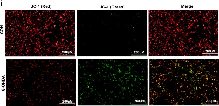
Same mechanism: at high potential it accumulates inside mitochondria, forms aggregates, and those emit red fluorescence, generally near 585 nm. At a fallen potential it stays monomeric mostly, and the green fluorescence comes out, generally near 527 nm.
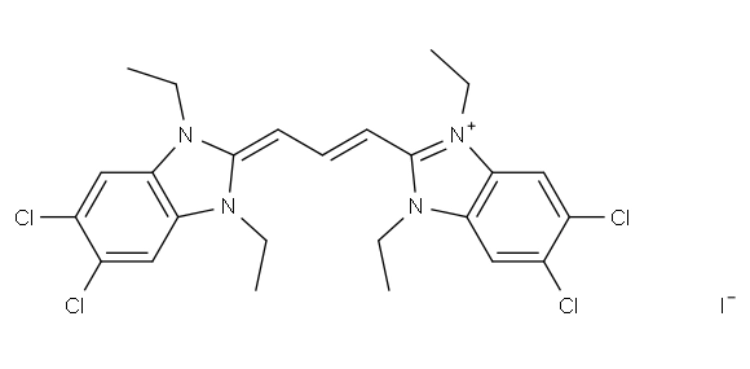
Read the red-to-green fluorescence ratio — that’s standard practice. Ratio comes down, membrane potential came down with it, usually.
Routine cell experiments, drug response studies, and apoptosis-related research all suit JC-1. The long history of use helps too, because your numbers can be lined up against a large body of previously published studies.
Solarbio JC-1 (Cat. No. IJ0300) has a reported purity of at least 95% by HPLC, and it can carry work that needs mitochondrial membrane potential changes monitored in cultured cells.
Rhodamine 123 and Its Green Signal
A cationic dye, Rhodamine 123. Mitochondria accumulate it according to mitochondrial membrane potential, and it fluoresces green, emission sitting around 527 nm.
With the potential intact, accumulation continues and nobody thinks about it. When potential decreases — well, mitochondrial accumulation and fluorescence distribution may change, and that’s your cue.
There’s no red-to-green aggregation transition in Rhodamine 123, nothing like JC-1 or JC-10. The information rides on fluorescence intensity and intracellular distribution instead; researchers watch those two to monitor mitochondrial changes.
Home ground, for it: cell viability studies, mitochondrial function research, drug-induced mitochondrial damage experiments.
Solarbio fournit Rhodamine 123 (Cat. No. IR1800), with a reported purity of at least 98%.
Mitochondrial Structure-Labeling Probes
MitoTracker Green, for Staining the Network
A fluorescent dye that labels mitochondria plainly enough that their distribution inside cells becomes visible — MitoTracker Green, most labs’ first call.
Green fluorescence, emission around 510 nm, good for following mitochondrial morphology, distribution, and shifts in the mitochondrial network.
People pull it into localization work and colocalization with other cellular structures as well. One caution, and it’s repeated on more forums than you’d think: don’t assume the staining behavior is completely independent of membrane potential. The specific formulation and your experimental conditions decide, so check both.
MitoTracker Red, the Red Channel Version
Red fluorescence is how MitoTracker Red labels mitochondria. Depending on the specific dye formulation, membrane potential may still pull on its mitochondrial uptake.
Standard use is distribution and morphology observation. Pair it with green fluorescent probes, though, and the relationship between mitochondria and other organelles opens up.
Mitochondrial autophagy research does exactly that — MitoTracker Red with LysoTracker Green, watching where mitochondria and lysosomes sit relative to each other.
Solarbio offers a Mitochondrial Red Fluorescent Probe (Cat. No. IF1770), with a reported purity of at least 98% and excitation/emission wavelengths of 579/599 nm. A Mitochondrial Green Fluorescent Probe (Cat. No. IF1780) is also available, with excitation/emission wavelengths of 490/523 nm.
Reported Experimental Conditions, and the Products Behind Them
Probe choice shifts with the cell model. The examples below are starting points for researchers planning mitochondrial fluorescence experiments.
JC-1 (Cat. No. IJ0300) has been used for fluorescence observation in 4T1 cells. One reported working condition is 5 μg/mL at 37°C and 5% CO2 for 20 minutes.
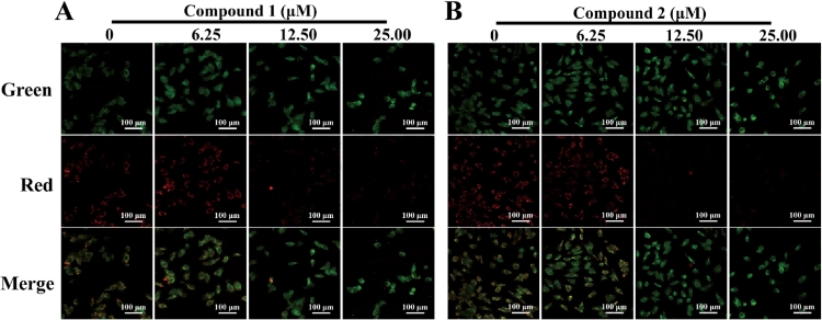
JC-10 (Cat. No. IJ0310) has seen use in RAW264.7 cell experiments observing mitochondrial membrane potential.
Rhodamine 123 (Cat. No. IR1800) has been used in SKOV3 and SKOV3TR cells. One reported experimental condition is 50 μg/mL, followed by incubation in the dark for 30 minutes.
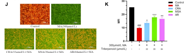
The Mitochondrial Red Fluorescent Probe (Cat. No. IF1770) has also been used for mitochondrial staining in HeLa cells.
Useful starting points, none of them universal protocols. For the cell type and setup in front of you, each of these gets optimized: probe concentration, incubation time, cell density, washing steps, imaging settings.
Researchers can explore related Solarbio research solutions for additional products and support across different life science applications.
Choosing Between These Probes Without Overthinking
One question runs the whole selection: what do you actually want to measure?
Detecting mitochondrial membrane potential changes is the goal — JC-1 and JC-10 both fit, and their red and green fluorescence signals make comparing different mitochondrial functional states possible.
Need the membrane potential-related fluorescence change carried on a green signal instead? Rhodamine 123 earns a look as the practical choice.
Morphology, distribution, or localization experiments belong with MitoTracker Green and MitoTracker Red, frankly.
Wavelengths are the next check. Excitation and emission against your fluorescence microscope or flow cytometer, because the probe must fit the available optical filters and shouldn’t overlap too much with other fluorescent labels in the same experiment.
For studies involving apoptosis, mitochondrial membrane potential results can be combined with other detection methods. Solarbio’s article on Méthodes de détection de l'apoptose : choix du test, conseils pratiques et kits recommandés explains how different methods can help researchers examine different stages of cell death.
Don’t forget the biological context, either. A change in mitochondrial fluorescence is not automatically a change in cell viability, and confirming results often takes additional markers. Related research topics sit in the Solarbio research pathways section.
Conclusion
Function, structure, cellular responses — mitochondrial fluorescent probes make all three visible somehow. Through red and green fluorescence signals, JC-1 and JC-10 read changes in mitochondrial membrane potential. Rhodamine 123 monitors membrane potential-related changes a different way. And for mitochondrial distribution and morphology, MitoTracker Green and MitoTracker Red are what most people reach for.
The best probe for every experiment doesn’t exist, however much we’d like it to. Research question, cell type, detection equipment, staining conditions — those decide. Match probe to purpose, optimize the protocol, and the fluorescence data holds up.
Solarbio provides a range of fluorescent probes and life science research products. Researchers can learn more about its technical and experimental services or visit the Solarbio company profile to learn more about its research product range and capabilities.
FAQ (questions fréquentes)
Q1: What do people do with mitochondrial fluorescent probes all day?
A1: Observe mitochondrial structure, distribution, and function. Most of the day’s work with them is monitoring mitochondrial membrane potential, investigating apoptosis, and following mitochondrial changes under different experimental conditions.
Q2: JC-1 versus JC-10 — since they work alike?
A2: Right, both are membrane potential-sensitive. Aggregate them at a higher mitochondrial membrane potential and they go red; at a lower one they sit as monomers and go green. Where they differ is staining performance and experimental behavior, so the cell type and protocol requirements pick the winner.
Q3: How does Rhodamine 123 actually read membrane potential?
A3: Positive charge, so mitochondria accumulate it according to the membrane potential. What gets measured is the shift — in mitochondrial accumulation and fluorescence intensity — as function changes.
Q4: MitoTracker Green vs MitoTracker Red, real difference?
A4: Both label mitochondria; the channels differ. Green goes green, Red goes red. Uptake and retention properties hang on the specific dye formulation, actually.
Q5: Any of them usable for live-cell imaging?
A5: Many are, yes. Product specifications, staining conditions, probe concentration, potential effects on cell viability — all four deserve a look before starting.
Q6: Can JC-1 serve as an apoptosis detector?
A6: Mitochondrial membrane potential loss, which is often associated with early apoptosis — JC-1 helps identify that. But the fluorescence change alone doesn’t confirm apoptosis, no. Pair JC-1 staining with something like Annexin V/PI staining or Caspase detection.
Q7: Walking into my first mitochondrial experiment — which probe?
A7: Decide what you want to observe, first thing. Membrane potential analysis wants JC-1 or JC-10. Membrane potential-related fluorescence changes suit Rhodamine 123. Mitochondrial labeling, MitoTracker Green or MitoTracker Red. Then make sure the fluorescence wavelengths suit your imaging equipment.
Product information, technical questions, cooperation inquiries: the Solarbio News Center handles those, or reach the Solarbio team directly.
 Consulter
 sur Whatsapp
 Laisser un message
 haut