






Related news
HDAC Explained: How Histone Deacetylases Control Gene Expression
Table of Contents
A mutation changes the function of a gene. This is well known to most researchers. However, the regulation of genes by a cell is much more complex, because genes can be up-regulated, down-regulated, or even completely repressed by the cell, all without any change of the DNA sequence.
Histone deacetylases, usually called HDACs, are involved in that control. They change the acetylation state of histones and many other proteins. The result may affect chromatin packing, transcription, cell growth, metabolism, immune signaling, and stress responses.
What Is HDAC and What Does It Do to Chromatin?
Histone Acetylation Loosens DNA Packing
DNA is not dissolved in the nucleus as free molecules but is packaged as chromatin, which is made up of DNA and histone proteins. The lysine residues on the histone tails can be acetylated by acetyl groups. The more acetyl groups that are added to the histone tails, the weaker the positive charge on these regions.
The bond between histones and negatively charged DNA then becomes less tight. Transcription factors and RNA polymerase have a better chance of reaching the gene.
Note that acetylation of histones does not activate all genes in the vicinity. Additional genes will be activated by their respective promoters, transcription factors, etc. in respective cell types. Additional chromatin proteins will also dominate over acetylated histones, opening up local areas of chromatin. It gives the transcription machinery access, but it does not decide the whole result by itself.
HDACs Remove Acetyl Groups
HDAC enzymes reverse this change. They remove acetyl groups from histone lysines. Once the acetyl group is gone, the lysine carries a stronger positive charge again. Histones can hold DNA more tightly, and the chromatin may become harder to access.
This is why HDACs are often linked with lower gene transcription. Some articles call them gene-silencing enzymes. That description is not completely wrong, but it is a little too simple.
An HDAC normally works with other proteins. Its effect depends on the complex it joins and the cell in which that complex is active. The same inhibitor can give one result in a tumor cell line and a different result in a primary immune cell. Looking only at the compound name is not enough.
Histones Are Not the Only Targets
HDACs also act on non-histone proteins. These include transcription factors, cytoskeletal proteins, molecular chaperones, and proteins involved in DNA repair. Removing an acetyl group may change a protein’s stability, location, binding partners, or activity.
HDAC6 is a good example. Much of its work is connected with cytoplasmic proteins such as alpha-tubulin. It is also involved in protein transport and protein quality control.
A researcher studying HDAC6 may learn very little by measuring histone acetylation alone. Acetylated alpha-tubulin, protein aggregation, cell movement, or related pathway markers may be more useful. Solarbio’s wider research product catalog includes compounds, antibodies, kits, and supporting reagents for these combined workflows.
How Are the Main HDAC Families Different?
Class I HDACs Are Closely Tied to Transcription
HDAC1, 2, 3, and 8 belong to Class I of the HDACs. They are found primarily in the nucleus and are well studied in terms of their role in transcription, as well as in cell-cycle control, proliferation, differentiation, and apoptosis.
HDAC1 and HDAC2 often sit inside large repressor complexes. HDAC3 also requires co-acting proteins to be fully active. It does not just roam around the nucleus in a non-specific fashion, removing acetyl groups.
This matters when reading an inhibitor result. A Class I inhibitor can potentially modulate the expression of a large number of genes. It may cause cells to enter a slow growth phase due to reasons such as cell-cycle arrest, apoptosis, or stress, and this slow growth phase can be due to multiple reasons. The effect of a histone acetylator on histone acetylation and the expression of target genes needs to be examined in more detail to draw a conclusion.
Class II and Class IV Work Differently
The class IIa HDACs are a subgroup of the class II histone deacetylases consisting of HDAC4, HDAC5, HDAC7, and HDAC9. These proteins are nuclear or cytoplasmic and can be highly mobile and transient between the two compartments, particularly in response to signaling events. These enzymes are relatively poor catalytic units but form stable multi-protein complexes with other proteins.
Class IIb are the HDAC6 and HDAC10 enzymes. More is known about HDAC6 because it is the only member of the HDAC family of enzymes to possess two catalytic domains. It is also known to be linked to the alpha-tubulin of the microtubules and to various steps of protein trafficking and protein homeostasis.
HDAC11 sits alone in Class IV. It is still being studied in immune regulation and metabolism. When a project focuses on one HDAC class instead of general deacetylase activity, it helps to check the related signaling pathways before choosing the inhibitor and downstream markers.
Sirtuins Use NAD+ Instead of Zinc
SIRT1 through SIRT7 are collectively known as the Class III HDACs. They are deacetylases that differ from the other HDACs in their mechanism of action and in the chemistry of their reaction. Most of the other HDACs are referred to as “zinc-dependent” because their active site contains a zinc ion that participates directly in the action of removing an acetyl group from a substrate. The sirtuins use NAD+ instead.
That link with NAD+ brings Sirtuins into studies on nutrient status, glucose and lipid metabolism, oxidative stress, mitochondria, and aging. Their location also varies. Some mainly work in the nucleus, while others are more closely linked with the cytoplasm or mitochondria.
A SIRT1 experiment should not simply copy a SIRT3 experiment. The substrates are different. The location is different. The readouts should be different as well. For SIRT3, mitochondrial acetylation and respiratory markers may be useful. For SIRT1, nuclear targets and metabolic signaling may deserve more attention.
How Does HDAC Catalysis Affect Inhibitor Choice?
Zinc-Dependent HDACs Share a Basic Reaction
Class I, Class II, and Class IV HDACs mainly use zinc in their catalytic pocket. The acetylated lysine enters the active site. Zinc helps position the acetyl group and a water molecule. Amino acids in the enzyme activate water, which then attacks the acetyl group.
Acetate is released, and the lysine returns to its deacetylated state.
Many HDAC inhibitors are designed around this pocket. One part of the molecule interacts with the zinc region. Other parts help the compound enter the channel and affect how well it fits different HDAC subtypes.
Two compounds may both carry the label “HDAC inhibitor,” yet behave very differently in cells. Their binding strength, subtype coverage, solubility, and cell permeability can all change the outcome.
Sirtuins Follow Another Chemical Route
Sirtuins do not use the same zinc-centered reaction. They consume NAD+ during deacetylation. In simple terms, NAD+ participates in catalytic reactions and undergoes decomposition. At the same time, it transfers the acetyl groups from histones to its own molecules, ultimately achieving the removal of the acetyl groups.The process produces nicotinamide and an ADP-ribose-related product.
Nicotinamide is often used in studies where researchers want to reduce Sirtuin activity. Resveratrol also appears in work related to Sirtuin signaling and metabolism. Neither compound should be treated as a perfect one-target switch.
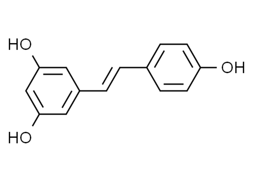
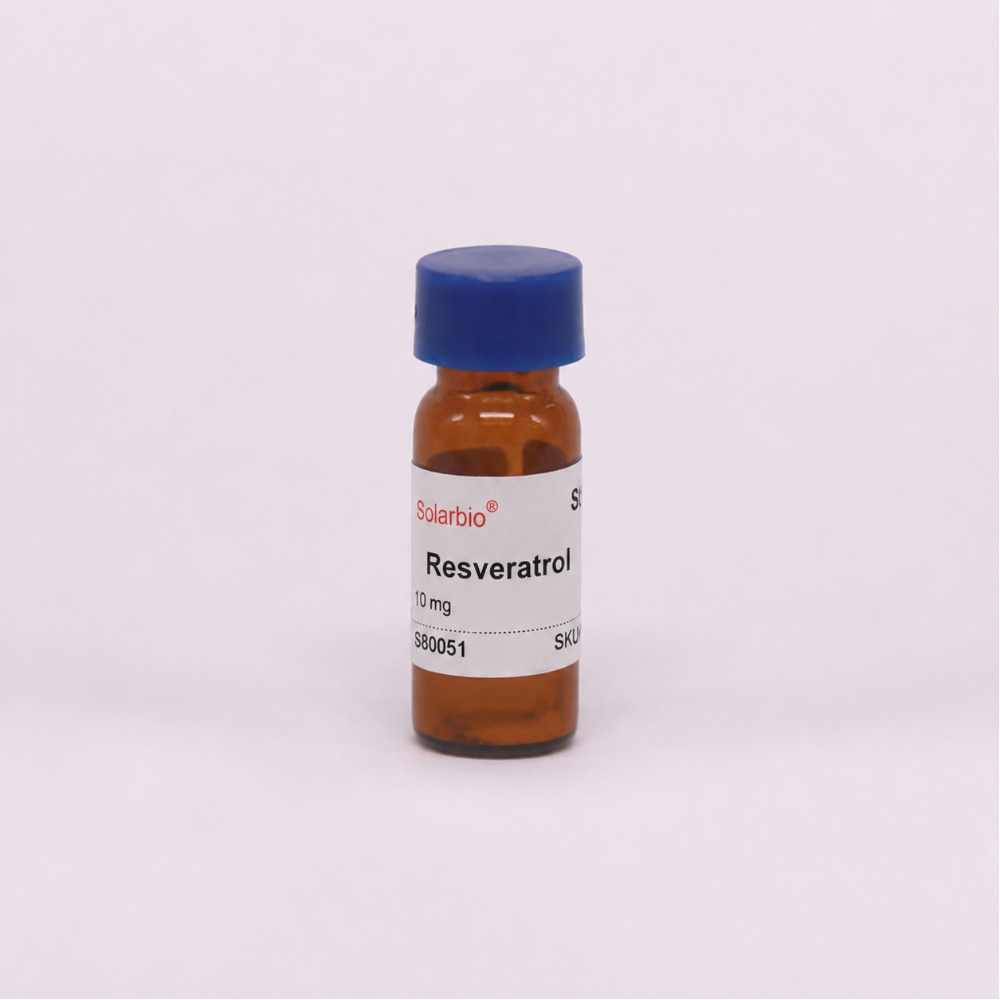
Cellular compounds often reach more than one protein. Dose also matters. A low concentration may give a fairly focused response, while a higher concentration brings in unrelated stress or off-target effects.
The safest approach is to pair the compound with a direct target marker or a known downstream response.
Selectivity depends on the working conditions
Vorinostat is commonly used as a broad HDAC inhibitor. Ricolinostat appears in many HDAC6-focused studies. TMP195 is often linked with Class IIa HDAC research.
Those descriptions are helpful when screening products, but they do not remove the need for validation. A compound that appears selective in a biochemical assay may become less selective when the cellular dose is raised.
Solvent level is another common problem. Many HDAC inhibitors are prepared in DMSO. When too much stock is added, the final DMSO concentration may affect membrane behavior, metabolism, or cell survival. The vehicle control must contain the same amount of solvent as every treated group.
Before the first plate is prepared, the concentration range, treatment time, vehicle level, and mechanism marker should already be clear.
Why Are HDACs Studied in Disease and Normal Cell Biology?
Cell Growth Depends on Controlled Gene Activity
HDACs are normal enzymes. Healthy cells need them. They help control when genes involved in growth, differentiation, and apoptosis are active.
During development, different tissues follow different gene programs. Liver cells, nerve cells, muscle cells, and immune cells do not use the same genes at the same time. HDAC family members help maintain those patterns.
Problems begin when the balance changes. In some cancer cells, high or misplaced HDAC activity may keep growth-control genes quiet. An inhibitor may help reopen those programs. It may also affect many other genes, which is why a drop in cell viability cannot explain the mechanism on its own.
Sirtuins Sit Close to Metabolic Changes
Sirtuins are often studied when researchers look at metabolism or mitochondria. Their dependence on NAD+ means their activity can shift with the cell’s energy and redox state.
SIRT1 is frequently linked with nuclear and metabolic signaling. SIRT3 is often examined through mitochondrial protein acetylation. Other Sirtuins have their own locations and functions.
A broad claim such as “the compound improves aging” is not very useful in the lab. It is better to ask what changed. Was it ATP production, mitochondrial membrane potential, reactive oxygen species, respiration, or a stress-response protein? Once the endpoint is clear, the experiment becomes easier to repeat.
Immune Responses Also Involve HDAC Activity
HDACs take part in T-cell activation, macrophage behavior, cytokine production, and inflammatory signaling. Both excessive and insufficient activity may disturb the balance.
The same inhibitor may reduce inflammatory markers in one model and harm cell survival in another. Primary cells are especially sensitive to donor differences, isolation methods, and activation state.
For projects that combine compounds, antibodies, cytokine assays, or several sample types, Solarbio’s technical services can help with assay selection and workflow planning. The aim is not to add more tests for the sake of it. Each test should answer a clear part of the research question.
How Can You Set Up a Cleaner HDAC Experiment?
Define the Target Before Buying the Compound
“Study HDAC inhibition” is not a strong experimental question. It is too open. A better question would be: does HDAC6 inhibition increase alpha-tubulin acetylation before it changes cell migration?
That sentence already tells you a lot. You need an HDAC6-related compound, an acetylated alpha-tubulin readout, at least two time points, and a migration assay.
The cell model should express the target. Check the baseline by qPCR or Western blotting, where possible. A negative result from a model with almost no target expression does not prove that the inhibitor failed.
Primary cells need another layer of care. Donor background, passage number, isolation conditions, and activation state may all change the response.
Use More Than One Readout
Cell viability is easy to measure, but it is not specific. A lower signal can come from apoptosis, cell-cycle arrest, metabolic suppression, membrane damage, or assay interference.
An HDAC experiment should include a marker closer to the target. For a Class I study, that may be histone acetylation and target gene expression. For HDAC6, acetylated alpha-tubulin is often more useful. For Sirtuins, mitochondrial or NAD+-related markers may fit better.
For general histone detection, the Anti-Histone H3.1 Monoclonal Antibody can support WB, IP, IF, IHC, or ELISA workflows according to the listed applications. A modification-specific antibody is still needed when the experiment focuses on a particular acetylation site.
Keep the controls simple and complete.
Start with a reasonable concentration range rather than one high dose. Include an early time point for acetylation and a later time point for gene expression or cell behavior.
The untreated control and vehicle control should both be present. When possible, add a known positive control. A second inhibitor with another chemical structure can help show whether the result follows the target rather than one compound’s off-target activity.
The cleanest data usually show an order. First, the acetylation marker changes. Then the pathway changes. The cell phenotype appears later.
Small handling details can still ruin that sequence. Repeated freeze-thaw cycles, the wrong dilution order, long room-temperature exposure, or uneven cell density can create a result that looks biological but is really technical. Solarbio’s technical articles and application notes can be checked before the first run for product handling and assay details.
Because an HDAC project is rarely about just buying one inhibitor and then measuring cell viability, you will most likely also need other antibodies, other pathway-modulating compounds, and a series of assays to monitor changes in apoptosis, release of cytokines, or in markers for mitochondria, to name but a few, before your data start to tell you a coherent story.
Beijing Solarbio Science & Technology Co., Ltd. has supplied life science research reagents since 2004. Its catalog covers small molecule compounds, antibodies, ELISA kits, biochemical assay kits, cell biology reagents, and related services. For researchers setting up work across several readouts, the company’s life science research solutions give a useful place to start.
Conclusion
HDACs are not a simple group of gene-silencing enzymes. Their work depends on the enzyme class, cellular location, catalytic mechanism, partner proteins, and the model used in the study.
A solid HDAC experiment starts with a clear target. The compound is then matched with a direct molecular readout, proper controls, and a later functional result. When those parts follow the same biological line, the data are easier to explain and much easier to repeat.
FAQ
Q1: What does HDAC mean?
A1: HDAC stands for Histone Deacetylase. These enzymes remove acetyl groups from histones and other proteins. This may change chromatin structure, protein behavior, and gene expression.
Q2: Why are HDACs important in epigenetics?
A2: HDACs can change gene activity without changing the DNA sequence. By removing acetyl groups from histones, they often make chromatin more compact and reduce access to certain genes.
Q3: Are all HDACs the same?
A3: No. HDACs are divided into several classes. Class I, Class II, and Class IV mainly use a zinc-dependent catalytic mechanism. Class III HDACs, known as Sirtuins, depend on NAD+.
 Consult
 Whatsapp
 Leave Message
 top
top