



Related news

Modified Sirius Red Staining Guide: Principle, Sample Choice, Troubleshooting, and Product Selection
Table of Contents
Collagen staining looks easy on paper, but anyone who has worked with tissue sections knows it can go wrong in small ways. The collagen signal may be weak. The background may turn red. The section may fall off. Sometimes the image under bright field looks acceptable, but the polarized light result does not show the fiber pattern clearly.
Modified Sirius Red staining is used often in fibrosis research, wound healing, skin repair, cardiac scar analysis, liver injury models, and extracellular matrix studies. It gives a clear collagen signal and, when observed under polarized light, helps show fiber arrangement and birefringence. That extra observation step is one reason many labs still keep this method in routine tissue staining work.
Solarbio provides staining reagents, biochemical reagents, molecular biology reagents, immunology products, and other research tools for life science research. For researchers using collagen staining, Modified Sirius Red staining is a very practical method for the following reasons: it is visual, it is direct and it can be correlated with the tissue morphology.
What Is Modified Sirius Red Staining?
A collagen-focused staining method for tissue sections
Modified Sirius Red staining is mainly used to stain collagen fibers in tissue sections. Sirius Red is a strong acidic dye. It binds to basic groups in collagen fibers, including groups related to lysine and hydroxyproline. After staining, collagen fibers usually appear red under bright-field microscopy.
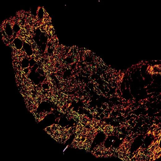
The useful part is polarized light observation. Collagen fibers can show birefringence. In common observation, type I collagen often appears orange-red, while type III collagen may show greenish tones. The exact color can still be affected by section thickness, staining time, tissue condition, and microscope settings, so a proper control slide is still needed.
For labs screening collagen staining tools, staining reagent products are a useful starting point for checking Sirius Red, Masson-related, Herovici, and other tissue staining options.
Why polarized light matters
Bright-field staining can show where collagen is located. Polarized light can provide more information about the collagen fibers, including their arrangement as well as the maturity of fibers. This property can be useful for the grading of fibrosis as well as for studies of tissue repair, of scar tissue, and for the study of connective tissue in general.
Modified Sirius Red staining does not replace molecular assays or protein-level detection. It cannot explain every mechanism behind collagen production. But for a slide-based view of collagen deposition, it is fast, readable, and practical.
Modified Sirius Red Staining vs Masson Trichrome Staining
The two methods are not the same
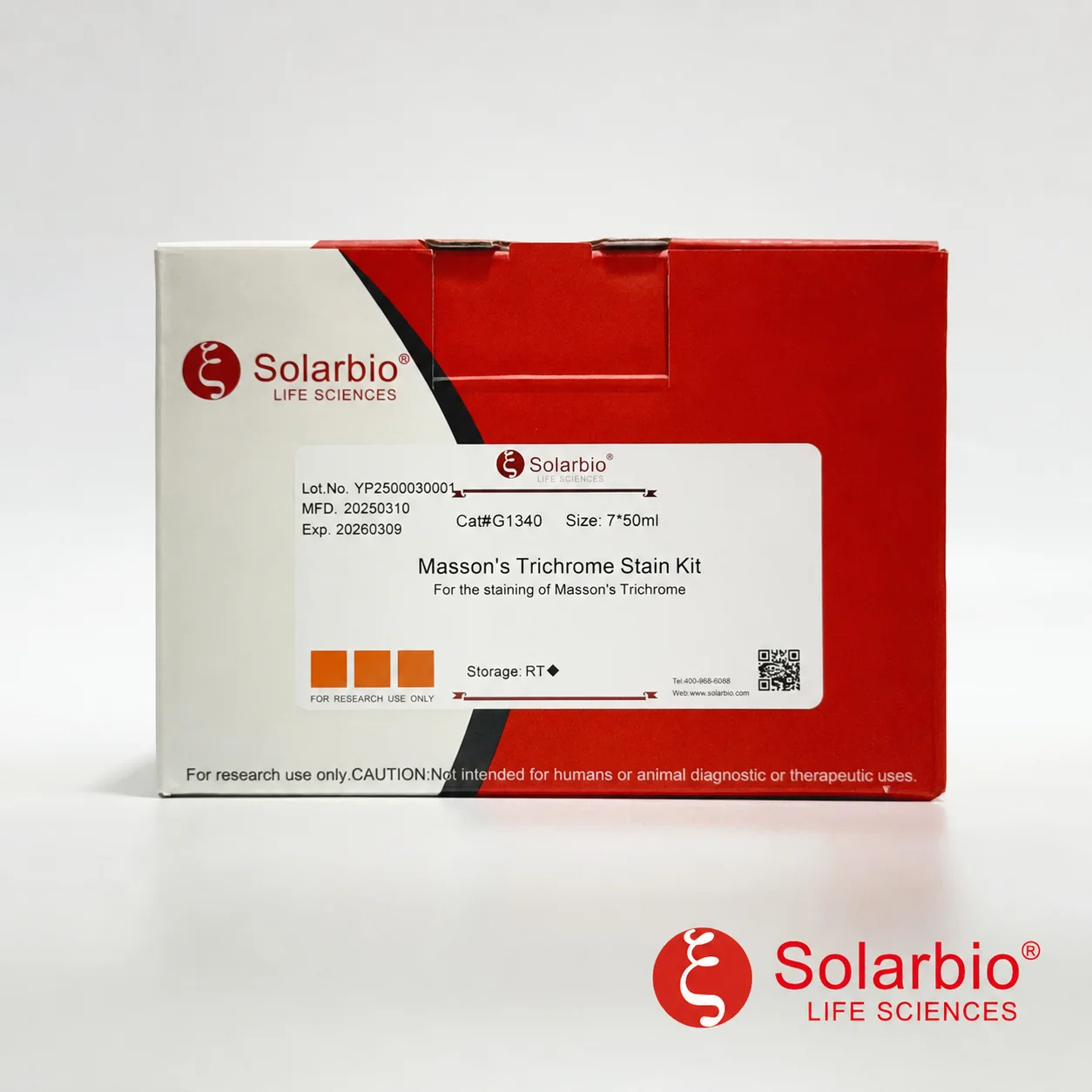
Masson Trichrome staining is also widely used for collagen observation. It gives a broader tissue structure view. The collagen in a section will usually be stained blue or green and the muscle and cytoplasm stained with a contrasting color depending on the particular kit or protocol used.
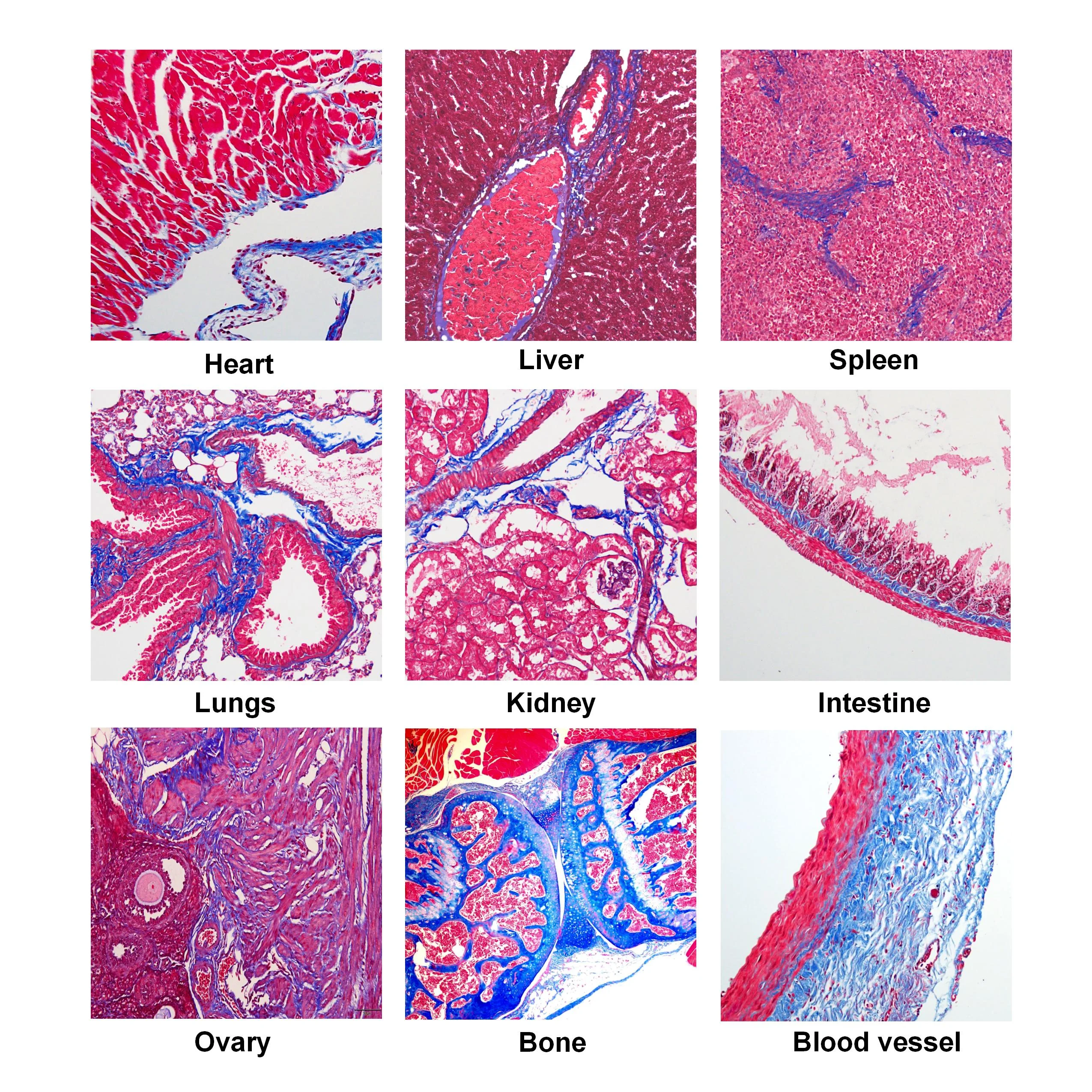
Modified Sirius Red staining is more focused on collagen fibers. Its value becomes stronger when polarized light is used. If the research question is about collagen fiber distribution and birefringence, Modified Sirius Red staining is often the better match. If the goal is to show collagen together with muscle, cytoplasm, and general tissue structure, Masson Trichrome staining may be easier to present.
|
Method |
Main Use |
Observation Advantage |
Suitable Sample |
|
Modified Sirius Red staining |
Collagen fiber staining |
Collagen birefringence under polarized light |
Paraffin and frozen tissue sections |
|
Masson Trichrome staining |
Collagen and general tissue structure |
Clear contrast between collagen and muscle or cytoplasm |
Paraffin and frozen tissue sections |
|
Herovici collagen staining |
Collagen maturity-related observation |
Helps compare young and mature collagen patterns |
Tissue sections |
|
Modified Aniline Blue staining |
Connective tissue staining |
Useful for collagen-related morphology |
Tissue sections |
For researchers comparing extracellular matrix methods, research pathways can help connect staining methods with broader fibrosis, tissue repair, and cell biology topics.
Which Samples Are Suitable?
Tissue sections are the right sample type
Modified Sirius Red staining is suitable for paraffin tissue sections and frozen tissue sections. It is not suitable for cultured cells, cell smears, or cell climbing slides. This point matters. Some failed staining results happen simply because the sample format was not right from the start.
For paraffin sections, 3–5 micrometers is commonly recommended, and 3 micrometers is often used for routine tissue sections. For myocardium or scar tissue, around 6 micrometers may work better because collagen distribution can be dense and uneven.
For frozen sections, 8–12 micrometers is often suggested. If the section falls off easily, reducing the thickness to around 7–8 micrometers may help.
Fixation and embedding notes
There is usually no special fixation requirement. Common fixatives such as 4% paraformaldehyde and 10% formalin can be used. Bouin’s fixative and Zenker fixative may also be used, but mercury-containing fixation needs proper mercury removal before staining.
Paraffin and frozen embedding are both acceptable. Plastic embedding, such as methyl methacrylate embedding, is not recommended because it can reduce dye penetration. When dye penetration is poor, the final collagen signal may look weak even if the staining reagent itself is working.
For labs building a complete histology workflow, pathological staining solutions may help when choosing between collagen staining, HE staining, Masson staining, and other tissue staining methods.
Product Selection for Collagen Staining
Main product options for collagen-related staining
The product group below covers Modified Sirius Red staining and several related connective tissue staining methods.
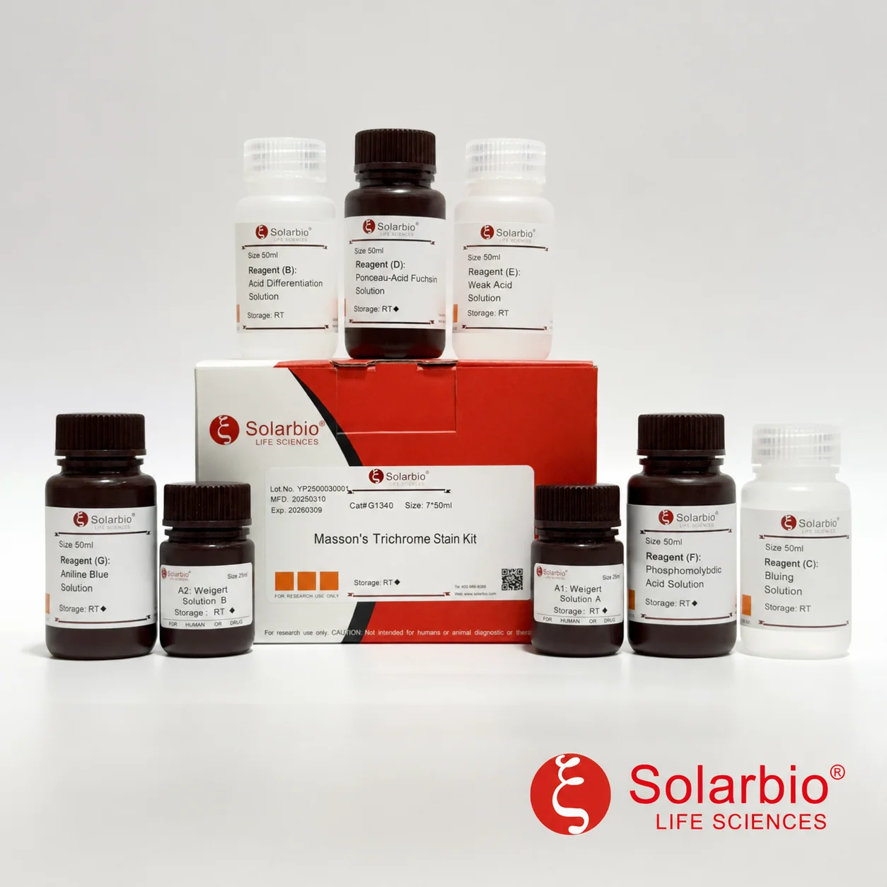
If the goal is direct Modified Sirius Red staining, G1472 Modified Sirius Red Staining Kit is the main kit option. If the lab only needs the core staining solution, G1473 Modified Sirius Red Staining Solution for Collagen Fiber Staining is the related staining solution.
|
Catalog No. |
Product Name |
Specification |
|
G1472 |
2 x 50 mL / 2 x 500 mL |
|
|
G1473 |
Modified Sirius Red Staining Solution for Collagen Fiber Staining |
100 mL / 500 mL |
|
G1475 |
3 x 50 mL / 3 x 100 mL |
|
|
G1476 |
2 x 50 mL / 2 x 500 mL |
|
|
G1340 |
7 x 50 mL / 7 x 100 mL |
|
|
G1343 |
Masson Trichrome Staining Kit, Fast Green Method |
7 x 50 mL / 7 x 100 mL |
|
G1346 |
8 x 50 mL / 8 x 100 mL |
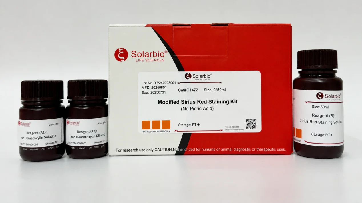
A simple way to choose is to start with the question. If the lab needs collagen fiber birefringence under polarized light, Modified Sirius Red staining is the better fit. If the lab needs a general tissue structure image with collagen contrast, Masson Trichrome staining may be more suitable. If collagen maturity is part of the research design, Herovici collagen staining can be considered.
Troubleshooting Common Staining Problems
Background is too red
A red background may come from overstaining, thick sections, insufficient washing, incomplete differentiation, or tissue preparation problems before staining. This is common, especially in tissue types that hold dye strongly.
The first adjustment should be simple. Shorten the staining time, improve washing, and check section thickness. Do not change too many conditions at the same time. If the section is too thick, background staining often becomes harder to control. For routine paraffin sections, starting around 3 micrometers is usually safer.
Collagen fibers are too pale
Weak collagen staining may come from short staining time, poor fixation, over-washing, poor dye penetration, or low collagen content in the sample itself. Another small but real issue is polarized light setup. If the microscope is not adjusted properly, the slide may look less clear than expected.
A positive control is useful here. A collagen-rich tissue, such as scar tissue or fibrotic tissue, helps decide whether the staining system is working. If the control is also weak, adjust staining time and washing conditions. If only the test sample is weak, the result may reflect the sample biology.
Nuclei appear red after staining
If Weigert’s iron hematoxylin is not used, light red nuclear staining may appear. This can be normal in the G1472 system. The kit uses a newer non-toxic yellow dye to replace picric acid. While it supports collagen and muscle fiber differentiation, it may also lead to some light nuclear staining. In most cases, this does not affect collagen judgment.
If nuclear staining needs to be clearer, Weigert’s iron hematoxylin can be added before Sirius Red staining.
Is iron hematoxylin required?
Iron hematoxylin is not required if nuclear staining is not needed. The section can go directly to Sirius Red staining. Routine hematoxylin, such as Mayer hematoxylin, is not recommended as a replacement. Weigert’s iron hematoxylin is acid-resistant and can better tolerate the acidic Sirius Red staining step. This helps reduce nuclear decolorization.
For labs that often meet staining problems, technical service support can help with product choice and basic troubleshooting before a full batch of valuable tissue sections is used.
Simple Checks Before Starting the Stain
Small checks reduce failed slides
Before staining a full batch, check the sample type first. It should be a paraffin or frozen tissue section, not a cultured cell sample. Then check thickness, fixation, slide adhesion, and the positive control.
A few practical checks are worth keeping:
Sample type should match the method.
Section thickness should match tissue type.
Fixation should be complete but not blocking dye penetration.
A collagen-rich positive control should be included.
Polarized light settings should be checked before final imaging.
Washing should be enough to reduce background but not so strong that weak collagen signal is lost.
These are ordinary steps, but they prevent many failed staining runs. The kit matters, of course. Still, the final slide also depends on section quality, fixation, timing, washing, and microscope settings.
For related product updates and method notes, users can check Solarbio news and technical articles.
Conclusion
Modified Sirius Red staining is a useful method for collagen fiber observation, especially when the stained section is checked under polarized light. It helps show collagen deposition, fiber distribution, and birefringence patterns in tissue sections.
The method works best when the sample type is correct. Paraffin and frozen tissue sections are suitable. Cultured cells are not. Section thickness, fixation, washing, staining time, and polarized light settings all affect the final image. When background turns too red or collagen looks weak, the first step should be checking the section and protocol conditions, not replacing everything at once.
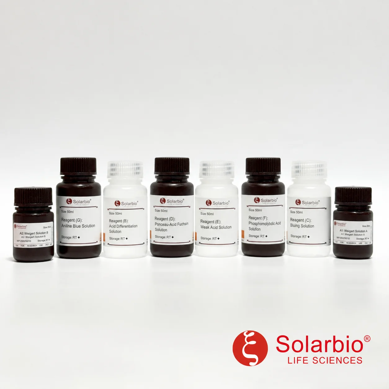
For product selection, G1472 Modified Sirius Red Staining Kit is the direct kit option. G1473 Modified Sirius Red Staining Solution for Collagen Fiber Staining, G1475 Herovici Collagen Staining Kit, G1476 Modified Aniline Blue Staining Kit, G1340 Masson Trichrome Staining Kit, G1343 Masson Trichrome Staining Kit, and G1346 Modified Masson Trichrome Staining Kit can be selected based on the staining goal.
Labs working on fibrosis, extracellular matrix, tissue repair, or collagen morphology can contact Solarbio with sample type, section thickness, fixation method, and observation needs.
FAQ
Q1: What is the reaction principle of Modified Sirius Red staining?
A1: Sirius Red is a strong acidic dye. It binds with basic groups in collagen fibers, including groups related to lysine and hydroxyproline. Under polarized light, collagen fibers show birefringence, which helps researchers observe collagen type and fiber arrangement more clearly.
Q2: What is G1472 Modified Sirius Red Staining Kit used for?
A2: G1472 Modified Sirius Red Staining Kit is used for collagen fiber staining in tissue sections. It is suitable for paraffin sections and frozen sections, and it can be used for collagen observation under bright-field and polarized light microscopy.
Q3: What is the difference between Modified Sirius Red staining and Masson Trichrome staining?
A3: Modified Sirius Red staining focuses more directly on collagen fibers and can show birefringence under polarized light. Masson Trichrome staining gives a broader tissue structure view, usually showing collagen and muscle or cytoplasm in different colors.
Q4: What samples are suitable for Modified Sirius Red staining?
A4: The method is suitable for paraffin tissue sections and frozen tissue sections. It is not suitable for cultured cells, cell smears, or cell climbing slides.
Q5: Does Modified Sirius Red staining require a special fixative?
A5: In most cases, no special fixative is required. Common fixatives such as 4% paraformaldehyde and 10% formalin can be used. Bouin’s fixative and Zenker fixative may also be used, but mercury-containing fixation needs proper mercury removal.
Q6: What section thickness is recommended?
A6: Paraffin sections are usually 3–5 micrometers, with 3 micrometers commonly used. Myocardium or scar tissue may use around 6 micrometers. Frozen sections are usually 8–12 micrometers. If sections detach easily, 7–8 micrometers can be tried.
Q7: Why does the background become too red after staining?
A7: A red background may come from overstaining, thick sections, weak washing, incomplete differentiation, or tissue preparation issues. Shortening staining time and improving washing are common first adjustments.
Q8: Why are collagen fibers pale or unclear?
A8: The reason may be short staining time, low collagen content, over-washing, poor fixation, poor dye penetration, or unsuitable polarized light settings. A collagen-rich positive control should be used to check whether the staining system works.
Q9: Is red nuclear staining normal if Weigert’s iron hematoxylin is not used?
A9: Yes. In the G1472 system, light nuclear staining can appear if Weigert’s iron hematoxylin is skipped. It usually does not affect collagen judgment. If clearer nuclei are needed, Weigert’s iron hematoxylin can be used before Sirius Red staining.
Q10: Can routine hematoxylin replace Weigert’s iron hematoxylin?
A10: It is not recommended. Weigert’s iron hematoxylin is more acid-resistant and can better tolerate the acidic Sirius Red staining step. Routine hematoxylin, such as Mayer hematoxylin, may decolorize more easily.
 Consult
 Whatsapp
 Leave Message
 top
top