



Related news
How to Detect, Remove, and Prevent Mycoplasma in Cell Culture
Table of Contents
Mycoplasma is one of those cell culture problems that can sit in the background for a long time. The medium may still look clear. Cells may remain attached. Nothing seems seriously wrong at first.
Then the strange results start.
Cells grow more slowly than before. Transfection efficiency drops for no clear reason. The medium turns yellow faster than usual. Some cells gather in uneven groups, while others detach. Two experiments carried out with the same protocol no longer give the same result.
Mycoplasma contamination may already be affecting the culture by that point.
These organisms are only around 50 to 300 nm in size, allowing them to pass through some filters used to retain common bacteria. Mycoplasma also lacks a cell wall, so routine penicillin-streptomycin treatment does not provide reliable protection.
A working control plan needs three parts. The laboratory has to detect contamination early, deal with infected cultures quickly, and reduce the chance of the same problem coming back. For laboratories using fluorescence-based screening, a complete kit can simplify staining, fixation, mounting, and microscopy preparation. Researchers setting up a wider contamination management process can also review available cell culture solutions when planning daily procedures.
Why Is Mycoplasma Easy to Miss?
The Culture May Still Look Normal
Severe contamination is easier to notice. Cells may become granular, grow in patches, detach from the flask, or stop dividing normally.
Low-level contamination is much less obvious. Cells may continue to proliferate whilst undergoing metabolic and gene expression changes. Mycoplasma utilize nutrients from culture medium and can interfere with amino acid metabolism, nucleotide synthesis, DNA replication, RNA production and protein expression.
This can create much frustration in the lab when an operator is continually adjusting parameters for transfection or switching sera or repeating an assay, not realizing that the problem actually lies with the cell stock.
Some projects are more sensitive than others. Cell signaling, drug screening, gene expression, cytokine analysis, and transfection work can all be affected. Researchers working with these applications may also check relevant cell biology pathways when reviewing unexpected changes in experimental results.
Routine Antibiotics Can Hide the Problem
Many laboratories add penicillin and streptomycin to the medium as a standard habit. These antibiotics help control some common bacteria, but they should not be treated as a mycoplasma prevention system.
Mycoplasma has no cell wall. Antibiotics that act on cell-wall formation therefore have little effect on it. The culture may remain clear while the contamination continues.
Long-term antibiotic use can also make the situation harder to notice. It may suppress other bacteria without removing mycoplasma, leaving the operator with a culture that looks clean but is not suitable for reliable experiments.
Where Does Cell Culture Mycoplasma Come From?
People Are a Common Source
A surprising amount of contamination starts with ordinary handling.
Mycoplasma may be carried in saliva and respiratory droplets. Talking over an open flask, touching a phone while wearing gloves, moving between doors and the biosafety cabinet, or keeping the same gloves on for too long can bring contaminants into the work area.
Spraying gloves does not make every surface contact disappear. Once gloves touch a chair, notebook, door handle, or phone, replacing them is usually safer.
Spilled medium is another common source. A small splash inside the cabinet can spread droplets onto pipettes, bottles, racks, and nearby culture vessels. It should be cleaned at once rather than left until the end of the session.
New Cells May Arrive Contaminated
Cells received from another laboratory should not go directly into the main culture room. A cell line may look healthy and still carry mycoplasma.
Primary tissue also carries more risk because it comes directly from animal or human material. Even careful sampling cannot remove every possible source of contamination.
New cells should be cultured separately, tested, and kept under quarantine until a negative result is confirmed. A clean stock can then be frozen for later use.
Shared Reagents Spread Contamination Quickly
A contaminated bottle of medium, PBS, trypsin, or serum can affect more than one cell line. The same applies to shared pipettes, aspiration tubing, and water baths.
The safest habit is simple. Handle one cell line at a time. Use small reagent aliquots. Keep quarantine reagents separate from routine culture reagents. Avoid carrying the same bottle between different work areas.
Which Mycoplasma Detection Method Works Best?
PCR and qPCR Are Practical for Routine Screening
PCR-based testing is widely used because it gives a clear result within a few hours. The method usually detects conserved mycoplasma DNA sequences, including regions associated with 16S rRNA genes.
It suits new cell screening, regular cell bank checks, troubleshooting, serum or medium evaluation, and pre-publication quality control.
Solarbio supplies several mycoplasma detection products for cell culture work. These include CA1081 Mycoplasma Test Kit (PCR Method) and CA1082 Mycoplasma Rapid Test Kit (for Cell Culture).
Hoechst Staining Is Useful for a Quick Check
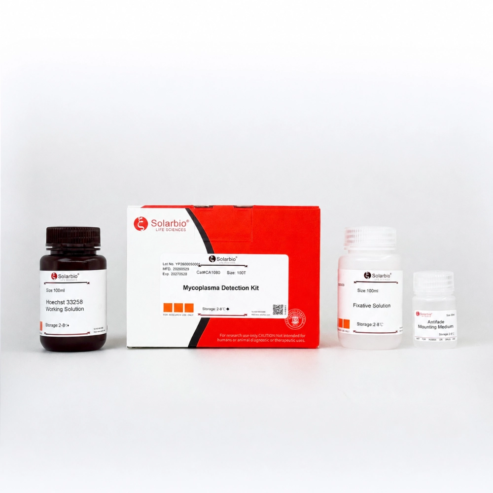
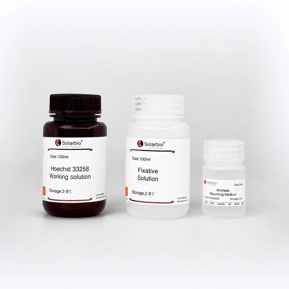
Hoechst fluorescent staining is cheap and does not require complicated equipment beyond a suitable fluorescence microscope. For fluorescence-based screening, the CA1080 Mycoplasma Detection Kit is designed for microscope-based mycoplasma checks in cell culture.
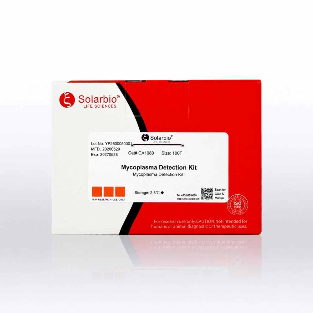
The kit includes Hoechst 33258 Working Solution, Fixative Solution, and Antifade Mounting Medium for staining, fixation, and slide preparation.
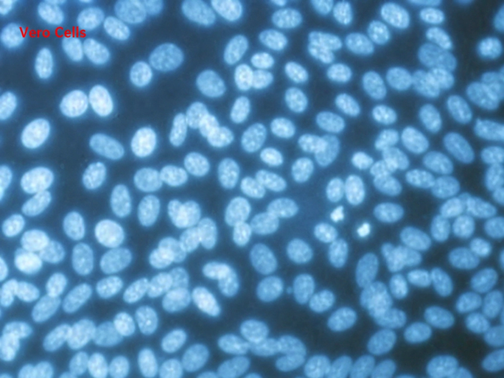
In a negative Vero cell sample, Hoechst fluorescence is mainly confined to the nuclei, without numerous fine fluorescent dots outside the nuclei.
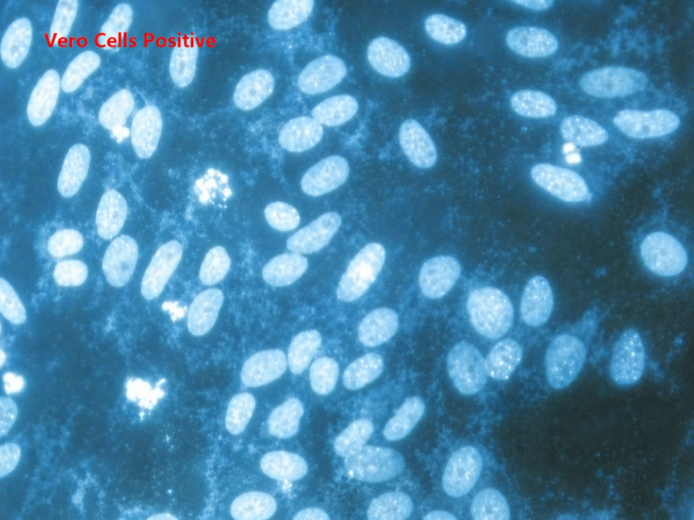
When contamination is present, small fluorescent dots may appear around the cell surface or between cells.
The test is useful for classroom work, quick checks, and laboratories with limited testing budgets.
It does have limits. Cell debris, apoptotic bodies, and background fluorescence may look similar to mycoplasma. A doubtful staining result should be confirmed by PCR.
Culture Testing Takes More Time
Culture-based testing remains important in some regulated settings and product release workflows. The sample is placed on suitable culture media and monitored for mycoplasma growth.
The main problem is speed. Results may take 7 to 21 days. This is not practical when a laboratory needs to decide quickly whether a cell batch can be used.
Some mycoplasma species are also harder to culture than others, so the method depends on suitable media and experienced operators.
Luminescence Tests Give Fast Results
Rapid luminescence assays detect enzyme activity or metabolic signals linked to viable mycoplasma. They are useful when many samples need to be screened in a short time.
The laboratory needs compatible equipment, and background interference should be checked carefully. For high-risk work, a rapid result may still need PCR or culture confirmation.
Teams that are not sure which method fits their cell type or testing schedule can use Solarbio’s technical service resources for product and workflow support.
Should a Contaminated Cell Line Be Treated?
Some Cultures Are Better Discarded
Treatment is not always the right choice.
If the cell line is easy to replace, heavily contaminated, or already stored as a clean frozen stock, disposal is normally safer. Keeping a badly contaminated culture in the laboratory creates a risk for every nearby cell line.
Rescue is more reasonable when the cells are rare, genetically edited, selected over a long period, derived from limited primary tissue, or difficult to obtain again.
The infected culture must be isolated before treatment begins. The incubator, cabinet, aspiration system, pipettes, water bath, and nearby surfaces should also be cleaned. Treating only the cells will not help much if the working area remains contaminated.
Homemade Antibiotic Treatment Is Hard to Control
Macrolides, tetracyclines, quinolones, and related compounds may work against some mycoplasma species. The difficult part is finding a concentration that removes the organism without killing the cells.
A dose that one cell line tolerates may stop another cell line from growing. Sensitive primary cells are especially difficult.
A low dose may only suppress contamination. The culture may test negative during treatment and become positive again later. For this reason, homemade antibiotic treatment should begin with a small tolerance test rather than the whole cell stock.
Commercial Removal Reagents Are Easier to Use
Commercial mycoplasma removal reagents save the laboratory from preparing several antibiotics and testing many concentrations from scratch.
Solarbio’s CA1090 MYCOPLASMA Scavenger and CA1093 Mycoplasma Elimination Reagent are intended for contaminated cell culture workflows. Treatment often continues through several passages, depending on the cell type and contamination level.
The cells should not be tested only while the removal reagent is still present. After treatment, they need a recovery period in reagent-free medium. Testing should then be repeated. A second negative result after further culture gives a more dependable answer.
How Can Mycoplasma Contamination Be Prevented?
Quarantine Cells Before Routine Use
Every incoming cell line should have a record. The record should include its source, arrival date, passage number, test result, operator, and storage position.
The culture should remain separate until testing is complete. After a negative result, a clean master stock should be frozen. Future work can then start from this stock instead of an old culture that has been passed for months.
Clean Shared Equipment Regularly
The biosafety cabinet needs cleaning before and after use. Incubator water trays should be emptied and disinfected on a regular schedule. Water baths, centrifuge carriers, microscope stages, aspiration bottles, and pipettes also need attention.
Solarbio CA1091 Mycoplasma Removal Spray can be used as part of an environmental cleaning process. CA1101 Mycoplasma Prevention Reagent is available for preventive cell culture use. Laboratories can check product updates when reviewing their contamination control materials.
Test Cells at the Right Time
Testing once a year is not enough for an active cell culture laboratory.
Cells should be tested when they arrive, before a master stock is frozen, after an important stock is thawed, before a long experiment, before cells are shared with another laboratory, and whenever unusual growth appears.
A three-month testing interval is a reasonable starting point for many research laboratories. High-throughput facilities or laboratories that regularly receive outside cell lines may need to test more often.
Good records matter as much as the test itself. When every cell line has a clear history, it becomes much easier to trace where contamination started and which cultures may have been exposed.
Solarbio supplies cell biology, molecular biology, immunology, biochemical, staining, analytical standard, and small molecule research products. Its management systems include ISO 9001, ISO 13485, ISO 14001, and ISO 45001.
Conclusion
Mycoplasma control does not need a complicated system, but it does need regular work.
Test new cells before they enter the main laboratory. Keep different cell lines and reagents separate. Clean shared equipment on schedule. Freeze a clean master stock while the culture is still in good condition.
When contamination is found, isolate the cells first. Replace the culture when possible. If the cells are valuable, use a controlled removal process and confirm the result after the treatment reagent has been removed.
The main point is not to wait for the medium to change color or for cells to start falling off the flask. By then, the experimental data may already have been affected.
For help choosing a suitable detection, removal, or prevention product, contact the Solarbio team.
FAQ
Q1: Can penicillin-streptomycin prevent mycoplasma contamination?
A1: No. Penicillin-streptomycin does not provide reliable protection because mycoplasma lacks the normal cell-wall structure targeted by many standard antibiotics.
Q2: How often should cell cultures be tested for mycoplasma?
A2: Many laboratories test every three months. Testing should also be carried out when cells arrive, before freezing a master stock, before major experiments, and when abnormal growth appears.
Q3: Can mycoplasma contamination be identified by looking at the medium?
A3: Not with confidence. The medium may remain clear during early contamination. Fast yellowing, slow cell growth, poor transfection, or unusual morphology may be warning signs, but a proper test is still needed.
Q4: Is PCR better than Hoechst staining for mycoplasma detection?
A4: The two methods can complement each other: Hoechst staining is well-suited for routine monitoring and preliminary screening, while PCR is more appropriate for confirmatory testing of suspected contamination or scenarios requiring higher sensitivity. For critical samples or biopharmaceutical lot release testing, it is recommended to combine culture-based methods (the gold standard) or employ multiple methods for cross-validation to enhance the reliability of the results.
Q5: Should every contaminated cell line be rescued?
A5: No. Replaceable or heavily contaminated cells are usually safer to discard. Rescue is mainly considered for rare, primary, selected, or genetically modified cell lines.
Q6: When should cells be retested after treatment?
A6: After mycoplasma‑elimination treatment, perform the first PCR test following 2‑3 passages (7‑10 days). Conduct the second test at a 7‑day interval. Two consecutive negative results indicate preliminary efficacy. Continuous monitoring until a negative result at day 28 confirms eradication.
Q7: Which Solarbio products cover mycoplasma detection and control?
A7: Solarbio provides CA1080, CA1081, and CA1082 for detection, CA1090 and CA1093 for removal, CA1091 for environmental cleaning, and CA1101 for prevention.
 Consult
 Whatsapp
 Leave Message
 top
top