





Related news
Human IL-23 ELISA Detection in Cell Culture Supernatants, Serum, and Plasma Samples
Table of Contents
IL-23 is a cytokine frequently measured in studies of inflammation, Th17 biology, immune activation, autoimmune disease, infection, and tumor immunity. Because IL-23 is a central component of the IL-23/IL-17 axis, it is often evaluated together with related mediators such as IL-17A, IL-22, and GM-CSF. Additional pathway markers may also be analyzed depending on the biological question.
The ELISA procedure itself is not necessarily complicated; however, pre-analytical and biological variables can strongly influence the result. Cell type, stimulation conditions, culture duration, sample handling and storage, and the expected IL-23 concentration can all affect the measured value. In healthy-donor serum or plasma, IL-23 may be below the assay’s detection capability, whereas appropriately stimulated IL-23-producing cell cultures can yield readily measurable concentrations.
The following examples involve peripheral blood cell preparations, THP-1 cells, Jurkat cells, serum, and plasma. They also illustrate an important point: the suitability of each model for IL-23 measurement is not equivalent, and results must be interpreted in the context of the cell type and stimulation conditions.
What Is Human IL-23?
IL-23 Is Made of Two Different Subunits
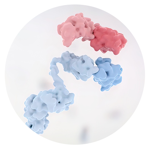
Human IL-23 belongs to the IL-12 cytokine family. It is a heterodimer rather than a single-chain protein.
IL-23 consists of a p19 subunit encoded by IL23A and a p40 subunit encoded by IL12B. The human p19 precursor is 189 amino acids long. The p40 subunit is shared with IL-12, and p19 and p40 associate as a disulfide-linked heterodimer to form biologically active IL-23.
This structure matters for detection. An assay intended to measure intact IL-23 should distinguish the p19/p40 heterodimer from related IL-12-family proteins and isolated subunits according to the assay’s validated specificity. Species and target specificity should therefore be confirmed before testing.
Solarbio supplies cytokine detection products and other immunology research solutions for cell-based and sample-based studies.
IL-23 Helps Maintain the Th17 Response
IL-23 signals through a heterodimeric receptor composed of IL-23R and IL-12Rβ1. This receptor is expressed on several responsive immune-cell populations, including activated and memory CD4+ T cells.
IL-23 receptor engagement activates JAK2/TYK2-associated signaling and predominantly STAT3, with STAT4 contributing to a lesser extent in many cellular contexts. This signaling supports the Th17 effector program, including RORγt expression and production of IL-17A, IL-22, and GM-CSF.
IL-23 is especially important for the maintenance, expansion, and pathogenic effector function of Th17 cells rather than acting as the sole initiator of naïve T-cell differentiation. This is why IL-23 is often measured in studies of chronic inflammation, autoimmune mechanisms, infection, and tumor immunity.
Researchers looking at the wider network can use Solarbio’s signaling pathway section to find related targets and pathway products.
How Should IL-23 Be Evaluated in Peripheral Blood Cell Cultures?
LPS Stimulation Requires an Appropriate Cell Preparation
For an LPS-induced IL-23 experiment, human peripheral blood mononuclear cells (PBMCs) or another preparation containing monocytes or other antigen-presenting cells is more appropriate than purified peripheral blood lymphocytes. IL-23 is produced predominantly by activated monocytes, macrophages, and dendritic cells, and LPS responses in human blood-cell preparations are largely dependent on innate LPS-sensing pathways. If the original material was recorded as “peripheral blood lymphocytes,” the isolation method and monocyte content should be verified before interpreting the result.
In the test setup described here, cells were cultured for 24 or 96 hours, and the culture supernatant was collected for human IL-23 measurement. If LPS concentrations of 50 ng/mL and 1 μg/mL are used, they should be treated as study-specific experimental conditions rather than universal recommended doses.
With two LPS concentrations and two collection times, the design yields four stimulated conditions. Comparisons should be made at matched time points and with an unstimulated control for each time point.
The findings should not be interpreted as a simple higher-dose-versus-lower-dose comparison. During a 96-hour culture, nutrient depletion, changes in cell density, cell death, shifts in cell composition, and cytokine degradation or consumption can all alter the measured concentration in the supernatant.
An unstimulated control should be maintained under identical culture conditions at each time point. Without this control, LPS-induced secretion cannot be distinguished reliably from basal secretion or time-dependent changes in the culture.
Culture Details Need to Stay Consistent
Small changes in culture handling can create large differences in cytokine measurements. Starting cell density, serum percentage, total culture volume, LPS preparation, incubation conditions, and collection method should be kept consistent across groups. For primary blood-cell experiments, donor source and the cell-isolation method should also be recorded because differences in monocyte content can strongly affect LPS-induced IL-23.
All supernatants should be processed consistently after collection. Avoid comparing a fresh sample with a sample that has undergone multiple freeze-thaw cycles.
Solarbio’s research product range covers ELISA kits, cell culture reagents, stimulants, and other products used in immune cell experiments.
How Does IL-23 Detection Work in THP-1 Cells?
A Monocytic Cell Model With LPS Stimulation
The human monocytic leukemia cell line THP-1 is widely used as a reproducible model for studies of innate immune signaling and inflammation. Primary human blood cells remain biologically important but show donor-to-donor variability and have a limited ex vivo lifespan; THP-1 cells can complement, but should not automatically replace, primary-cell experiments.
In this example, THP-1 cells were cultured in RPMI-1640 medium with 10% fetal bovine serum. LPS was added at final concentrations of 50 ng/mL and 500 ng/mL.
Culture supernatants were collected after 24 and 48 hours and then tested for human IL-23.
The two LPS concentrations should not automatically be labeled as “moderate” and “strong.” THP-1 responsiveness to LPS depends on cell state, differentiation status, cell density, serum conditions, and receptor expression; a higher LPS concentration does not necessarily produce a proportionally higher or more interpretable IL-23 response.
THP-1 Results Should Not Be Treated Like Primary Cell Results
THP-1 cells and primary human monocytes or PBMCs do not have the same origin or biological behavior. Their receptor expression, activation state, growth pattern, and response to LPS can differ.
A result from THP-1 cells should first be interpreted within the THP-1 model. It should not be used as a direct substitute for data from primary human immune cells.
When the assay signal is weak, the standard curve looks unusual, or repeated runs do not agree, Solarbio’s technical service team can help review product choice and basic test conditions.
Are Jurkat Cells an Appropriate Model for IL-23 Secretion?
PMA Stimulation Requires Cautious Interpretation
Jurkat cells are a human T-cell leukemia line widely used for studies of T-cell signaling. However, activated monocytes, macrophages, and dendritic cells are the principal established cellular sources of secreted heterodimeric IL-23, so Jurkat cells are not a standard positive-production model for IL-23.
In the setup described here, PMA was used at final concentrations of 0.2 μg/mL and 10 μg/mL for three or five days before supernatant collection. These are strong and prolonged stimulation conditions for Jurkat cells and should not be presented as generally recommended IL-23-induction conditions. Any IL-23 signal obtained under these conditions should be interpreted together with unstimulated controls and cell-viability data.
The large concentration range and multi-day exposure make a simple dose-response interpretation difficult, because changes in viability, proliferation, and cell state may dominate the measured outcome.
Cell viability and cell number should be monitored throughout the experiment, particularly at the higher PMA concentration and after prolonged exposure. A detectable ELISA signal alone does not establish that Jurkat cells are a physiologically appropriate IL-23-production model; unexpected positive results should be confirmed with appropriate controls and, when necessary, an orthogonal method.
The Stimulant and Cell Type Must Match the Study Goal
LPS and PMA are not interchangeable stimulants. LPS activates TLR4-dependent signaling in responsive innate immune cells, whereas PMA directly activates protein kinase C-dependent pathways and is commonly used to trigger signaling in several cell models.
The purpose of the experiment should determine the cell model and stimulus. THP-1 cells or primary monocyte-lineage cells may be used to study innate responses to LPS. Jurkat cells with PMA can be useful for T-cell signaling studies, but this does not make Jurkat cells a validated model of IL-23 secretion. The ELISA result must be interpreted within the biology of the chosen model.
Solarbio publishes laboratory articles and product updates that cover ELISA detection, cell experiments, and common testing issues.
Why Can Normal Serum and Plasma Show No Detectable IL-23?
Results May Fall Below the Detection Limit
In this sample panel, 20 normal human plasma samples and 20 normal human serum samples were tested, and IL-23 was below the assay’s limit of detection in all samples.
This finding does not prove that IL-23 was completely absent. It means that, under the assay conditions used, any IL-23 present was below the assay’s stated detection capability.
Many cytokines, including IL-23, may circulate at very low concentrations in healthy individuals. Measured concentrations can also be influenced by immune status, inflammation or infection, medication, age, sample handling, and other biological or pre-analytical factors.
A result below the limit of detection is not automatically an assay failure. The calibration curve, blank and control performance, sample handling, and assay procedure should be reviewed first. If assay performance is acceptable, the result should be reported as below the detection limit, or according to the manufacturer’s specified reporting convention, rather than assigned an exact concentration.
Serum and Plasma Are Different Matrices
Serum and plasma should not automatically be combined into a single analytical group. Their preparation methods differ, and matrix effects can influence assay performance.
The collection tube, anticoagulant used for plasma, clotting conditions for serum, time to centrifugation, storage temperature, and number of freeze-thaw cycles should all be documented. These variables become especially important when the target concentration is near the lower end of the assay’s reportable range.
Which IL-23 ELISA Kit Fits the Sample?
Human IL-23 ELISA Kit
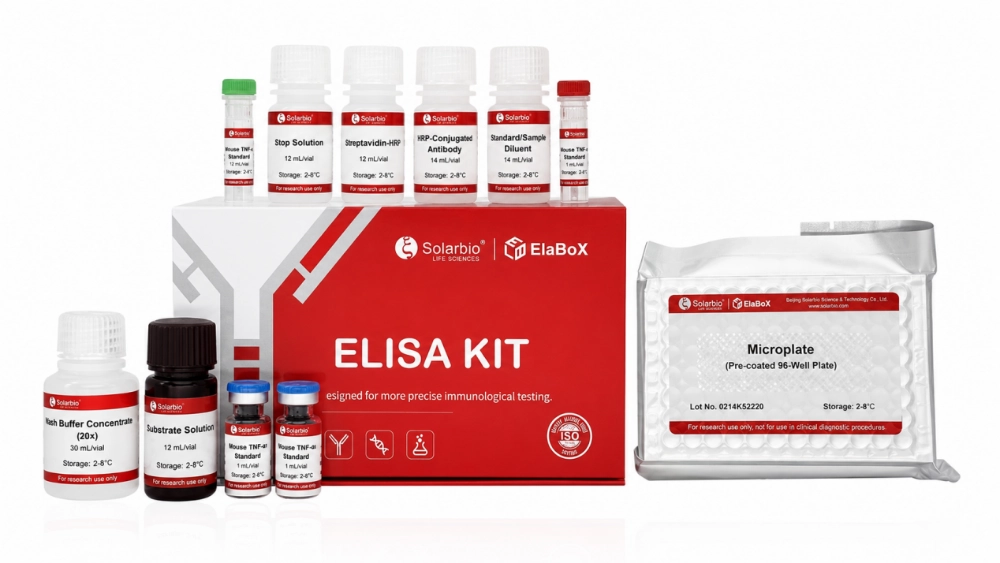
The SEKH-0034 Human IL-23 ELISA Kit can be used for the quantitative detection of human IL-23 in validated human sample types, including serum, plasma, and cell culture supernatants, in accordance with the current product instructions.
The product is relevant to inflammation research, Th17-related studies, immune cell stimulation, autoimmune mechanisms, infection, and tumor immunity.
The SEKH-0034 Human IL-23 ELISA Kit should be used with sample matrices validated in the current product instructions. Before testing, the expected concentration should also be considered so that samples fall within, or can be brought into, the assay’s reportable range using a validated dilution procedure.
IL-23 Kits for Other Species
SEKM-0024 Mouse IL-23 ELISA Kit is intended for mouse samples. SEKR-0072 Rat IL-23 ELISA Kit is used for rat samples, and SEKCN-0130 Chicken IL-23 ELISA Kit is available for chicken research.
A kit made for one species should not be used for another species unless cross-reactivity has been tested and confirmed. Similar protein names do not mean the assay antibodies will recognize the target in the same way.
The Mouse IL-23 ELISA Kit and other species-specific kits should be selected according to the animal model used in the study.
Beijing Solarbio Science & Technology Co., Ltd. was founded in 2004 and develops products for immunology, cell biology, molecular biology, and biochemistry. More information about its product and quality system is available on the Solarbio company page.
What Should Be Checked Before Reading the Final Result?
Start With the Standard Curve
The calibration curve should meet the kit’s stated acceptance criteria and use the recommended curve-fitting approach. Replicate wells should agree within the assay’s specified precision limits.
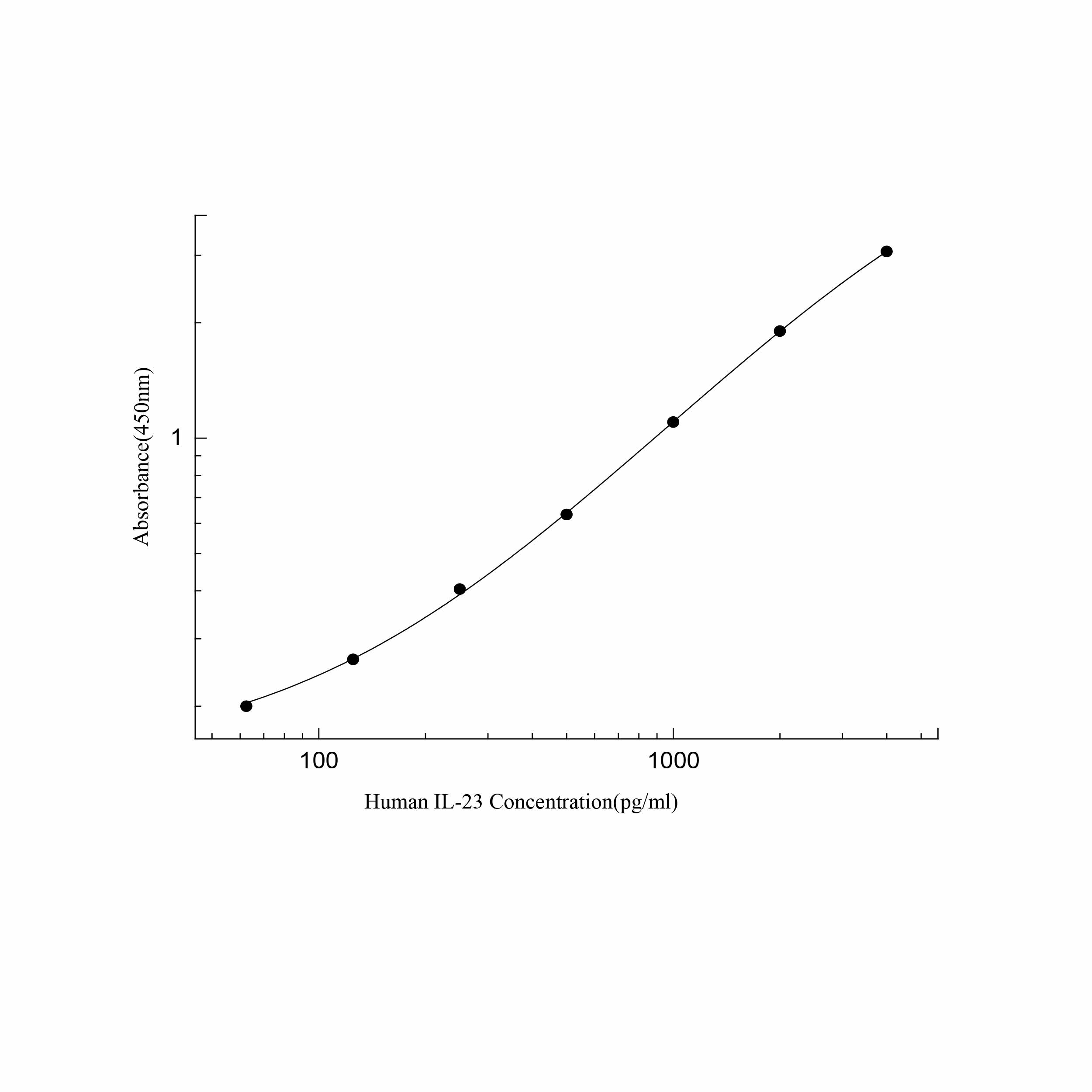
Blank absorbance should remain low. A high blank may point to incomplete washing, contamination, incorrect reagent handling, or excessive incubation.
Samples with signals above the assay’s upper reportable limit should be diluted and retested if the protocol allows. Results below the lower limit of quantification should not be reported as exact concentrations, and results below the limit of detection should be reported accordingly.
Look Back at the Sample Preparation
Unexpected IL-23 values often lead investigators to question the kit first, but the source of the discrepancy may have occurred earlier in the workflow.
Cell density may have differed between groups, the stimulant may have been prepared incorrectly, or the supernatant may contain cell debris. Serum or plasma samples may also have undergone repeated freeze-thaw cycles.
Pipetting, washing, incubation time and temperature, and microplate-reader settings also need to be consistent. ELISA is sensitive to handling differences, especially when the cytokine concentration is low.
Conclusion
Human IL-23 measurement can provide useful information in inflammation and immune-response studies, but the measured concentration must be interpreted in the context of the full experimental design.
For LPS-induced IL-23 studies, monocyte-containing PBMCs, primary monocytes or dendritic cells, and appropriately characterized myeloid cell models are biologically more suitable than purified lymphocytes. THP-1 cells can be evaluated with LPS under validated culture conditions. Jurkat cells may be useful for PMA-driven T-cell signaling studies, but they should not be presented as a standard IL-23 secretion model without supporting validation.
Normal serum and plasma may show IL-23 levels below the detection limit. This can be a valid result when the assay controls and standard curve perform normally.
Species matching, sample handling, culture consistency, and assay execution all affect the final data. For questions about kit selection or test conditions, researchers can contact Solarbio before starting the assay.
FAQ
Q1: What is Human IL-23?
A1: Human IL-23 is a pro-inflammatory heterodimeric cytokine in the IL-12 family. It consists of the IL-23A-encoded p19 subunit and the IL-12B-encoded p40 subunit.
Q2: Why is IL-23 linked to Th17 cells?
A2: IL-23 signals through a receptor complex composed of IL-23R and IL-12Rβ1. This activates JAK2/TYK2-associated signaling, with STAT3 as the predominant downstream STAT in Th17-related responses.
Q3: Can Human IL-23 be detected in cell culture supernatants?
A3: Yes. Cell-culture supernatants can be tested when the cell model is capable of producing IL-23 and the sample type is validated for the ELISA kit. Monocyte-containing PBMCs, primary monocytes/macrophages/dendritic cells, and suitably characterized myeloid cell models are generally more appropriate for IL-23 secretion studies than purified lymphocytes or Jurkat cells.
Q4: Can LPS be used in an IL-23 experiment?
A4: Yes. LPS can stimulate IL-23 production in responsive innate immune cells, including monocytes, macrophages, dendritic cells, monocyte-containing PBMC preparations, and some myeloid cell models. It should not be assumed to induce IL-23 secretion from purified lymphocytes.
Q5: Can PMA be used with Jurkat cells?
A5: PMA can activate Jurkat cells and is useful in T-cell signaling studies, but Jurkat cells are not a standard positive-production model for secreted IL-23. PMA-treated Jurkat supernatants should not be presented as a validated IL-23 positive control without supporting data.
Q6: Why is IL-23 sometimes undetectable in normal serum or plasma?
A6: IL-23 may be present at a concentration below the assay detection limit. This is common with low-abundance cytokines in normal samples.
Q7: Can a Mouse IL-23 ELISA Kit test human samples?
A7: No. Human samples should be tested with a Human IL-23 ELISA Kit unless the manufacturer has specifically validated cross-reactivity.
Q8: What should be checked when duplicate wells do not match?
A8: Pipetting accuracy, sample mixing, washing, incubation time, plate position, bubbles, and reagent preparation should all be reviewed.
 Consult
 Whatsapp
 Leave Message
 top
top